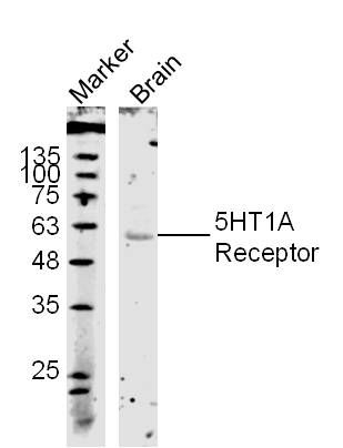
Protein: Brain(Mouse) Lysate at 40 ug
Primary: Rabbit Anti- 5HT1A Receptor (SL1124R) at 1:300;
Secondary: 800CW Conjugated Goat (polyclonal) Anti-Rabbit IgG(H+L) at 1: 10000;
Predicted band size:46 kD
Observed band size:56 kD
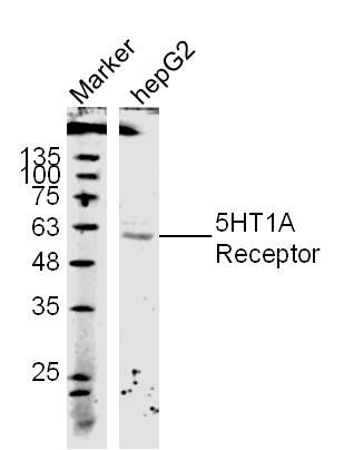
Protein: HepG2 lyates(hu);
Primary: Rabbit Anti- 5HT1A Receptor (SL1124R) at 1:300;
Secondary: 800CW Conjugated Goat (polyclonal) Anti-Rabbit IgG(H+L) at 1: 10000;
Predicted band size:46 kD
Observed band size:56 kD
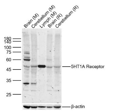
Sample:
Lane 1: Mouse Brain Lysates
Lane 2: Mouse Cerebellum Lysates
Lane 3: Mouse Lymph Lysates
Lane 4: Rat Brain Lysates
Lane 5: Rat Cerebellum Lysates
Primary: Anti-5HT1A Receptor (SL1124R) at 1/1000 dilution
Secondary: IRDye800CW Goat Anti-Rabbit IgG at 1/20000 dilution
Predicted band size: 46kDa
Observed band size: 46kDa
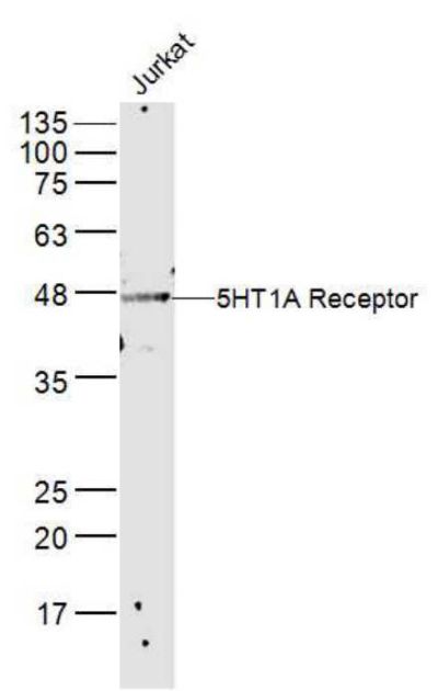
Sample:
Jurkat(Human) Cell Lysate at 40 ug
Primary: Anti-5HT1A Receptor (SL1124R) at 1/300 dilution
Secondary: IRDye800CW Goat Anti-Rabbit IgG at 1/20000 dilution
Predicted band size: 46 kD
Observed band size: 46 kD
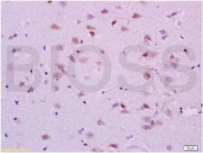
Tissue/cell: rat brain; 4% Paraformaldehyde-fixed and paraffin-embedded;
Antigen retrieval: citrate buffer ( 0.01M, pH 6.0 ), Boiling bathing for 15min; Block endogenous peroxidase by 3% Hydrogen peroxide for 30min; Blocking buffer (normal goat serum,SLC0005) at 37℃ for 20 min;
Incubation: Anti-5HT1A Receptor Polyclonal Antibody, Unconjugated(SL1124R) 1:400, overnight at 4°C, followed by conjugation to the secondary antibody(SP-0023) and DAB(SLC0010) staining
|