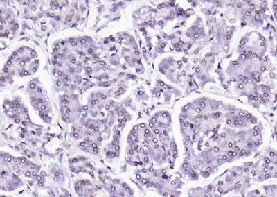
Paraformaldehyde-fixed, paraffin embedded (human pancreatic cancer); Antigen retrieval by boiling in sodium citrate buffer (pH6.0) for 15min; Block endogenous peroxidase by 3% hydrogen peroxide for 20 minutes; Blocking buffer (normal goat serum) at 37°C for 30min; Antibody incubation with (VEGFR3) Polyclonal Antibody, Unconjugated (bSL2202R) at 1:200 overnight at 4°C, followed by operating according to SP Kit(Rabbit) (sp-0023) instructionsand DAB staining.
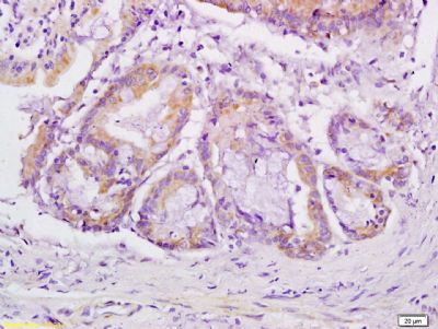
Tissue/cell: human gastric carcinoma;4% Paraformaldehyde-fixed and paraffin-embedded
Antigen retrieval: citrate buffer ( 0.01M, pH 6.0 ), Boiling bathing for 15min; Block endogenous peroxidase by 3% Hydrogen peroxide for 30min; Blocking buffer (normal goat serum) at 37℃ for 20 min
Incubation: Anti-VEGFR3 Polyclonal Antibody, Unconjugated(SL2202R) 1:200, overnight at 4°C, followed by conjugation to the secondary antibody and DAB staining
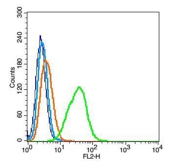
Blank control: A549(blue), the cells were fixed with 2% paraformaldehyde (10 min) and then permeabilized with ice-cold 90% methanol for 30 min on ice..
Isotype Control Antibody: Rabbit IgG(orange) ;
Secondary Antibody: Goat anti-rabbit IgG-FITC(white blue),
Dilution: 1:100 in 1 X PBS containing 0.5% BSA ;
Primary Antibody Dilution: 1μl in 100 μL1X PBS containing 0.5% BSA(green).
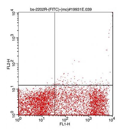
Cell: (mo)Splenocyte(2% BSA at 4°blocked for 30 minutes.1% Paraformaldehyde fixed for 30 minutes,0.1%TritonX-100 permeablized for 2 minutes).
Concentration:1:100.
Incubation: 40 minutes.
Host/Blank: Splenocytes.
Flow cytometric analysis of Rabbit Anti-VEGFR3 antibody (SL2202R)(green) compared with control in the absence of primary antibody (blue) followed by Splenocytes.
Secondary antibody: Goat Anti-rabbit IgG/FITC antibody (SL0295G-FITC).
|