Home > Product > Antibody > Rabbit Anti-Adenosine A3 Receptor antibody
adenosine A3 receptor; A3AR; AD 026; AD026; ADORA 3; ADORA3; bA552M11.5; RP11 552M11.7; TGPCR1; adenosine receptor A3 isoform 1; adenosine receptor A3 isoform 2; AA3R_HUMAN.
Cat:
SL1225R
Species Reactivity:
Human,Mouse,Rat,(predicted: Dog,Pig,Cow,Rabbit,Sheep,)
Immunogen:
KLH conjugated synthetic peptide derived from human ADORA3:85-180/347
Format:
Liquid
Storage instructions:
Shipped at 4℃. Store at -20 °C for one year. Avoid repeated freeze/thaw cycles.
Concentration:
1mg/ml
Clonality:
Polyclonal
Isotype:
IgG
Applications:
WB=1:500-2000ELISA=1:5000-10000IHC-P=1:100-500IHC-F=1:100-500Flow-Cyt=1μg /testIF=1:100-500(Paraffin sections need to do antigen repair)not yet tested in other applications.optimal dilutions/concentrations should be determined by the end user.
Host:
Rabbit
Product Overview:
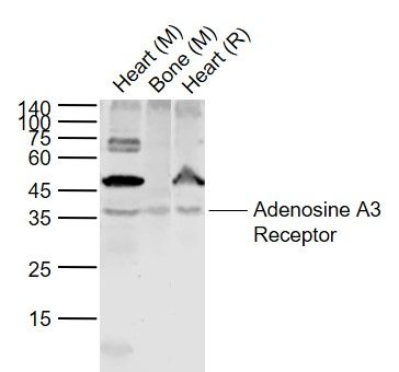
Sample: Lane 1: Heart (Mouse) Tissue Lysate at 40 ugLane 2: Bone (Mouse) Tissue Lysate at 40 ugLane 3: Heart (Rat) Tissue Lysate at 40 ugPrimary: Anti-Adenosine A3 Receptor (SL1225R) at 1/1000 dilutionSecondary: IRDye800CW Goat Anti-Rabbit IgG at 1/20000 dilutionPredicted band size: 35 kDObserved band size: 36 kDTissue/cell: human gastric carcinoma; 4% Paraformaldehyde-fixed and paraffin-embedded; Antigen retrieval: citrate buffer ( 0.01M, pH 6.0 ), Boiling bathing for 15min; Block endogenous peroxidase by 3% Hydrogen peroxide for 30min; Blocking buffer (normal goat serum,SLC0005) at 37℃ for 20 min; Incubation: Anti-ADORA3 Polyclonal Antibody, Unconjugated(SL1225R) 1:200, overnight at 4°C, followed by conjugation to the secondary antibody(SP-0023) and DAB(SLC0010) stainingParaformaldehyde-fixed, paraffin embedded (rat brain); Antigen retrieval by boiling in sodium citrate buffer (pH6.0) for 15min; Block endogenous peroxidase by 3% hydrogen peroxide for 20 minutes; Blocking buffer (normal goat serum) at 37°C for 30min; Antibody incubation with (Adenosine A3 Receptor) Polyclonal Antibody, Unconjugated (SL1225R) at 1:500 overnight at 4°C, followed by a conjugated secondary (sp-0023) for 20 minutes and DAB staining. Blank control (blue line): Raji (blue)(The cells were fixed with 70% ethanol overnight at -20℃). Primary Antibody (green line): Rabbit Anti-Adenosine A3 Receptor antibody(SL1225R); Dilution: 1μg /10^6 cells; Isotype Control Antibody (orange line): Rabbit IgG . Secondary Antibody (white blue line): Goat anti-rabbit IgG-PE;Dilution: 1μg /test. Blank control (blue line): Raji (blue)(The cells were fixed with 70% ethanol overnight at -20℃). Primary Antibody (green line): Rabbit Anti-Adenosine A3 Receptor antibody(SL1225R); Dilution: 1μg /10^6 cells; Isotype Control Antibody (orange line): Rabbit IgG . Secondary Antibody (white blue line): Goat anti-rabbit IgG-PE;Dilution: 1μg /test. Blank control:HL-60. Primary Antibody (green line): Rabbit Anti-Adenosine A3 Receptor antibody (SL1225R) Dilution: 2μg /10^6 cells; Isotype Control Antibody (orange line): Rabbit IgG . Secondary Antibody : Goat anti-rabbit IgG-FITCDilution: 1μg /test. ProtocolThe cells were incubated in 5%BSA to block non-specific protein-protein interactions for 30 min at room temperature .Cells stained with Primary Antibody for 30 min at room temperature. The secondary antibody used for 40 min at room temperature. Acquisition of 20,000 events was performed.
More
Unit:
Price: $
Product PDFs
Datasheet:


This gene encodes a protein that belongs to the family of adenosine receptors, which are G-protein-coupled receptors that are involved in a variety of intracellular signaling pathways and physiological functions. The receptor encoded by this gene mediates a sustained cardioprotective function during cardiac ischemia, it is involved in the inhibition of neutrophil degranulation in neutrophil-mediated tissue injury, it has been implicated in both neuroprotective and neurodegenerative effects, and it may also mediate both cell proliferation and cell death. Multiple transcript variants encoding different isoforms have been found for this gene. [provided by RefSeq, Jul 2008].

Function:
Receptor for adenosine. The activity of this receptor is mediated by G proteins which inhibits adenylyl cyclase. Possible role in reproduction.

Subcellular Location:
Cell membrane.

Post-translational modifications:
Phosphorylation on Ser-317 may be crucial for rapid desensitization.

Similarity:
Belongs to the G-protein coupled receptor 1 family.

SWISS:
P0DMS8

Gene ID:
140

Database links:

Entrez Gene: 403805 Dog

Entrez Gene: 140 Human

Entrez Gene: 606753 Pig

Omim: 600445 Human

SwissProt: Q28309 Dog

SwissProt: P0DMS8 Human


ADORA-3(腺苷受体A3)属于G蛋白偶联受体家族,在腺苷调节细胞活力和生长中起关键作用,并且在很多肿瘤中表达增高,A3AR可能成为肿瘤诊断标志物和治疗靶点。 Picture

Sample:
Lane 1: Heart (Mouse) Tissue Lysate at 40 ug
Lane 2: Bone (Mouse) Tissue Lysate at 40 ug
Lane 3: Heart (Rat) Tissue Lysate at 40 ug
Primary:
Anti-Adenosine A3 Receptor (SL1225R) at 1/1000 dilution
Secondary: IRDye800CW Goat Anti-Rabbit IgG at 1/20000 dilution
Predicted band size: 35 kD
Observed band size: 36 kD
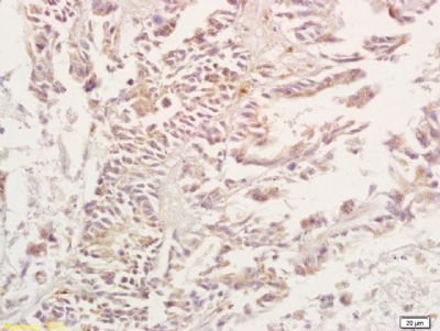
Tissue/cell: human gastric carcinoma; 4% Paraformaldehyde-fixed and paraffin-embedded;
Antigen retrieval: citrate buffer ( 0.01M, pH 6.0 ), Boiling bathing for 15min; Block endogenous peroxidase by 3% Hydrogen peroxide for 30min; Blocking buffer (normal goat serum,SLC0005) at 37℃ for 20 min;
Incubation: Anti-ADORA3 Polyclonal Antibody, Unconjugated(SL1225R) 1:200, overnight at 4°C, followed by conjugation to the secondary antibody(SP-0023) and DAB(SLC0010) staining
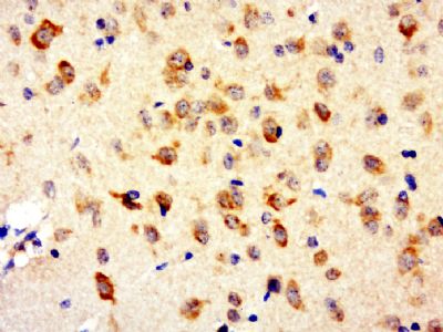
Paraformaldehyde-fixed, paraffin embedded (rat brain); Antigen retrieval by boiling in sodium citrate buffer (pH6.0) for 15min; Block endogenous peroxidase by 3% hydrogen peroxide for 20 minutes; Blocking buffer (normal goat serum) at 37°C for 30min; Antibody incubation with (Adenosine A3 Receptor) Polyclonal Antibody, Unconjugated (SL1225R) at 1:500 overnight at 4°C, followed by a conjugated secondary (sp-0023) for 20 minutes and DAB staining.
Blank control (blue line): Raji (blue)(The cells were fixed with 70% ethanol overnight at -20℃).
Primary Antibody (green line): Rabbit Anti-Adenosine A3 Receptor antibody(SL1225R); Dilution: 1μg /10^6 cells;
Isotype Control Antibody (orange line): Rabbit IgG .
Secondary Antibody (white blue line): Goat anti-rabbit IgG-PE;Dilution: 1μg /test.
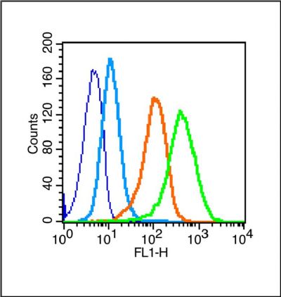
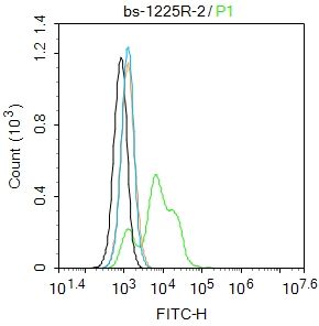
Blank control:HL-60.
Primary Antibody (green line): Rabbit Anti-Adenosine A3 Receptor antibody (SL1225R)
Dilution: 2μg /10^6 cells;
Isotype Control Antibody (orange line): Rabbit IgG .
Secondary Antibody : Goat anti-rabbit IgG-FITC
Dilution: 1μg /test.
Protocol
The cells were incubated in 5%BSA to block non-specific protein-protein interactions for 30 min at room temperature .Cells stained with Primary Antibody for 30 min at room temperature. The secondary antibody used for 40 min at room temperature. Acquisition of 20,000 events was performed.
Product Feedback Wall
Message :
Your Email :
Copyright © 2007-2018 Sunlong Medical All Rights Reserved.