[IF=2.245] Taotao Li. et al. Integrating miRNA and mRNA Profiling to Assess the Potential miRNA–mRNA Modules Linked With Testicular Immune Homeostasis in Sheep. Front Vet Sci. 2021; 8: 647153 WB,IF ; Sheep.
[IF=4.344] Gitto SB et al. Identification of a novel IL-5 signaling pathway in chronic pancreatitis and crosstalk with pancreatic tumor cells. Cell Commun Signal. 2020 Jun 17;18(1):95. IHSLCP ; Mouse.
[IF=3.943] Yihan Lu et al. Experimental evidence for alpha enolase as one potential autoantigen in the pathogenesis of both autoimmune thyroiditis and its related encephalopathy. Int Immunopharmacol. 2020 Aug;85:106563. IF ; Mouse.
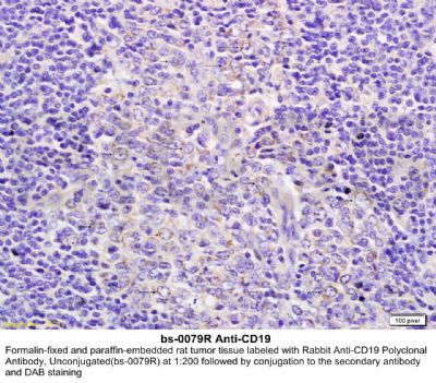
[IF=2.515] Li T et al. Digital gene expression analyses of mammary glands from meat ewes naturally infected with clinical mastitis.R Soc Open Sci. 2019 Jul 3;6(7):181604. IHC ; Sheep.
[IF=4.1] Goodwin, Justin, et al. "Targeting HIF-1α/PDK1 Axis by Dichloroacetate (DCA) Suppresses Bleomycin-induced Pulmonary Fibrosis." American Journal of Respiratory Cell and Molecular Biology ja (2017). IHSLCP ; Mouse.
[IF=0.52] Utomo, Pamudji, et al. "Decreasing SDF1-CXCR4 Expression after Adipose-Derived Mesenchymal Stem Cells (ASCS) Treatment Combined with Freeze-Dried Amniotic Membrane Wrapping in Rat Sciatic Nerve Injury." International Journal of ChemTech Research. IF(ICC) ; Rat.
[IF=1.813] Othman Hemn. et al. Antileukemic Effect of Palladium Nanoparticles Mediated by White Tea (Camellia sinensis) Extract In Vitro and in WEHI-3B-Induced Leukemia In Vivo. Evid-Based Compl Alt. 2020;2020:8712896 IHC ; Mouse.
[IF=2.08] Takahashi, Yuichi, Yohei Ikezumi, and Akihiko Saitoh. "Rituximab protects podocytes and exerts anti‐proteinuric effects in rat adriamycin‐induced nephropathy independent of B‐lymphocytes." Nephrology (2016). Rat.
[IF=3.54] Reese, Shannon R., et al. "Calcineurin Inhibitor Minimization With Ixazomib, an Investigational Proteasome Inhibitor, for the Prevention of Antibody Mediated Rejection in a Preclinical Model." Transplantation (2015). IHSLCP ; Rat.