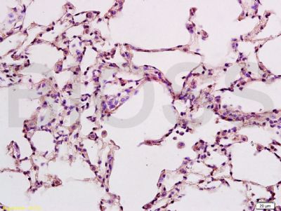
Tissue/cell: Mouse placenta tissue; 4% Paraformaldehyde-fixed and paraffin-embedded;
Antigen retrieval: citrate buffer ( 0.01M, pH 6.0 ), Boiling bathing for 15min; Block endogenous peroxidase by 3% Hydrogen peroxide for 30min; Blocking buffer (normal goat serum,SLC0005) at 37∩ for 20 min;
Incubation: Anti-CD274 Polyclonal Antibody, Unconjugated(SL1103R) 1:500, overnight at 4∑C, followed by conjugation to the secondary antibody(SP-0023) and DAB(SLC0010) staining
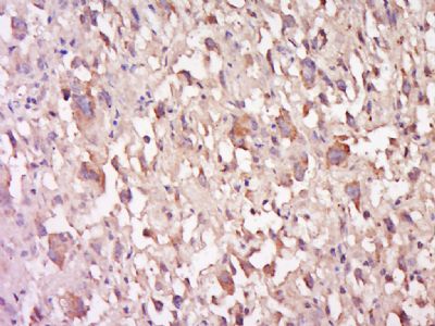
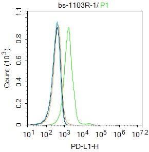
Blank control: Hela.
Primary Antibody (green line): Rabbit Anti-PD-L1 antibody (SL1103R)
Dilution: 1ug/Test;
Secondary Antibody : Goat anti-rabbit IgG-FITC
Dilution: 0.5ug/Test.
Protocol
The cells were incubated in 5%BSA to block non-specific protein-protein interactions for 30 min at room temperature .Cells stained with Primary Antibody for 30 min at room temperature. The secondary antibody used for 40 min at room temperature. Acquisition of 20,000 events was performed.