| Picture |
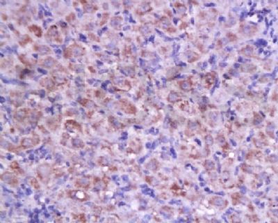
Tissue/cell: rat ovary tissue; 4% Paraformaldehyde-fixed and paraffin-embedded;
Antigen retrieval: citrate buffer ( 0.01M, pH 6.0 ), Boiling bathing for 15min; Block endogenous peroxidase by 3% Hydrogen peroxide for 30min; Blocking buffer (normal goat serum,SLC0005) at 37℃ for 20 min;
Incubation: Anti-Cyclin D2 Polyclonal Antibody, Unconjugated(SL1148R) 1:200, overnight at 4°C, followed by conjugation to the secondary antibody(SP-0023) and DAB(SLC0010) staining
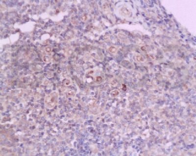
Tissue/cell: human flat moss tinea tissue; 4% Paraformaldehyde-fixed and paraffin-embedded;
Antigen retrieval: citrate buffer ( 0.01M, pH 6.0 ), Boiling bathing for 15min; Block endogenous peroxidase by 3% Hydrogen peroxide for 30min; Blocking buffer (normal goat serum,SLC0005) at 37℃ for 20 min;
Incubation: Anti-Cyclin D2 Polyclonal Antibody, Unconjugated(SL1148R) 1:200, overnight at 4°C, followed by conjugation to the secondary antibody(SP-0023) and DAB(SLC0010) staining
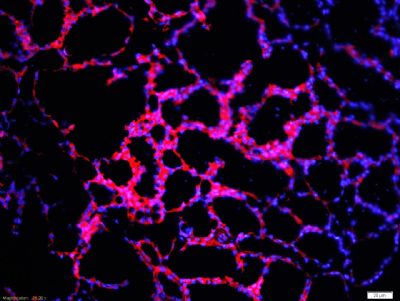
Tissue/cell: rat mammary tissue;4% Paraformaldehyde-fixed and paraffin-embedded;
Antigen retrieval: citrate buffer ( 0.01M, pH 6.0 ), Boiling bathing for 15min; Blocking buffer (normal goat serum,SLC0005) at 37℃ for 20 min;
Incubation: Anti-Cyclin D2 Polyclonal Antibody, Unconjugated(SL1148R) 1:200, overnight at 4°C; The secondary antibody was Goat Anti-Rabbit IgG, PE conjugated (SL0295G-PE)used at 1:200 dilution for 40 minutes at 37°C. DAPI(5ug/ml,blue,SLC0033) was used to stain the cell nuclei
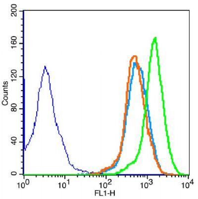
The figure annotation:
The blue histogram is unstained cells.
The Wathet Blue histogram is cells stained with secondary antibody(SL0295G-FITC) alone.
The Orange histogram is cells stained with rabbit IgG isotype control(SL0295P) antibody plus secondary antibody.
The green histogram is cells stained with Rabbit Anti-Cyclin D2 antibody (SL1148R)plus secondary antibody.
.
Concebtration: 5μg/10^6 cells.
Positive control: MCF-7 cells.
|