Neurofascin is a cell adhesion molecule involved in mediating axon recognition but also signaling axonal contact. Immunoglobulin domain cell adhesion molecule (cam) subfamily; members are components of neural cell adhesion molecules (N-CAM L1), Fasciclin II and the insect immune protein Hemolin. The subfamily also includes receptor domains such as as the extracelluar ligand binding domain of Fibroblast Growth Factor Receptor 2. Members are phylogenetically diverse, occuring throughout metazoa, and are not components of the adaptive immune system molecules found in jawed vertebrates. A predominant feature of most Ig domains is a disulfide bridge connecting 2 beta-sheets with a Trp packing against the disulfide bond.
Function:
Cell adhesion, ankyrin-binding protein which may be involved in neurite extension, axonal guidance, synaptogenesis, myelination and neuron-glial cell interactions.
Subunit:
Horseshoe-shaped homodimer.
Subcellular Location:
Cell membrane; Single-pass type I membrane protein.
Similarity:
Belongs to the immunoglobulin superfamily. L1/neurofascin/NgCAM family.
SWISS:
O94856
Gene ID:
23114
Database links:
Entrez Gene: 23114 Human
Entrez Gene: 269116 Mouse
Entrez Gene: 116690 Rat
Omim: 609145 Human
SwissProt: O94856 Human
SwissProt: Q810U3 Mouse
SwissProt: P97685 Rat
Unigene: 13349 Human
Unigene: 326702 Mouse
Unigene: 3048 Rat
神经生物学相关蛋白(Neurobiology)
| Picture |
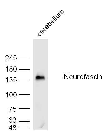
Sample: Cerebellum (Mouse) Lysate at 30 ug
Primary: Anti- Neurofascin Polyclonal (SL0289R) at 1/300 dilution
Secondary: IRDye800CW Goat Anti-Rabbit IgG at 1/20000 dilution
Predicted band size: 132/150 kD
Observed band size: 136 kD
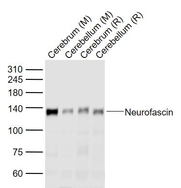
Sample:
Lane 1: Cerebrum (Mouse) Lysate at 40 ug
Lane 2: Cerebellum (Mouse) Lysate at 40 ug
Lane 3: Cerebrum (Rat) Lysate at 40 ug
Lane 4: Cerebellum (Rat) Lysate at 40 ug
Primary: Anti-Neurofascin (SL0289R) at 1/1000 dilution
Secondary: IRDye800CW Goat Anti-Rabbit IgG at 1/20000 dilution
Predicted band size: 132 kD
Observed band size: 132 kD
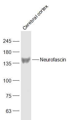
Sample:
Cerebral cortex (Mouse) Lysate at 40 ug
Primary: Anti-Neurofascin (SL0289R) at 1/1000 dilution
Secondary: IRDye800CW Goat Anti-Rabbit IgG at 1/20000 dilution
Predicted band size: 132/150 kD
Observed band size: 150 kD
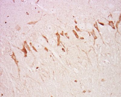
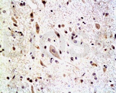
Tissue/cell: Rat brain tissue; 4% Paraformaldehyde-fixed and paraffin-embedded;
Antigen retrieval: citrate buffer ( 0.01M, pH 6.0 ), Boiling bathing for 15min; Block endogenous peroxidase by 3% Hydrogen peroxide for 30min; Blocking buffer (normal goat serum,SLC0005) at 37∩ for 20 min;
Incubation: Anti-Neurofascin Polyclonal Antibody, Unconjugated(SL0289R) 1:500, overnight at 4∑C, followed by conjugation to the secondary antibody(SP-0023) and DAB(SLC0010) staining
Tissue/cell: rat spinal cord tissue; 4% Paraformaldehyde-fixed and paraffin-embedded;
Antigen retrieval: citrate buffer ( 0.01M, pH 6.0 ), Boiling bathing for 15min; Block endogenous peroxidase by 3% Hydrogen peroxide for 30min; Blocking buffer (normal goat serum,SLC0005) at 37∩ for 20 min;
Incubation: Anti-Neurofascin Polyclonal Antibody, Unconjugated(SL0289R) 1:200, overnight at 4∑C, followed by conjugation to the secondary antibody(SP-0023) and DAB(SLC0010) staining
|
|
|