Home > Product > Antibody > Rabbit Anti-NM23A antibody
AWD; AWD, drosophila, homolog of; GAAD; Granzyme A activated DNase; Granzyme A-activated DNase; GZMA activated DNase; Metastasis inhibition factor NM23; NB; NBS; NDK A; NDKA; NDKA_HUMAN; NDP kinase A; NDPK-A; NDPKA; NM23; NM23 long variant
Cat:
SL1066R
Species Reactivity:
Human,Mouse,Rat,(predicted: Dog,Pig,Cow,Horse,Rabbit,)
Immunogen:
KLH conjugated synthetic peptide derived from human Nm23-H1:41-152/152
Format:
Liquid
Storage instructions:
Shipped at 4℃. Store at -20 °C for one year. Avoid repeated freeze/thaw cycles.
Concentration:
1mg/ml
Clonality:
Polyclonal
Isotype:
IgG
Applications:
WB=1:500-2000ELISA=1:5000-10000IHC-P=1:100-500IHC-F=1:100-500Flow-Cyt=1μg/Test IF=1:100-500(Paraffin sections need to do antigen repair)not yet tested in other applications.optimal dilutions/concentrations should be determined by the end user.
Host:
Rabbit
Product Overview:
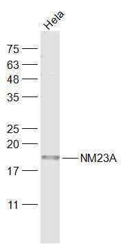
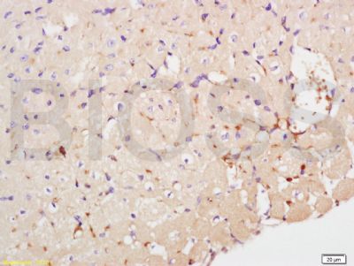
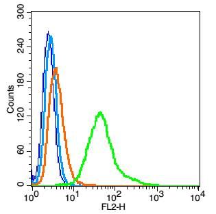
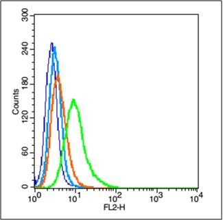
Sample: Hela(Human) Cell Lysate at 30 ugPrimary: Anti-NM23A (SL1066R) at 1/1000 dilutionSecondary: IRDye800CW Goat Anti-Rabbit IgG at 1/20000 dilutionPredicted band size: 17 kDObserved band size: 18 kDTissue/cell: mouse heart tissue; 4% Paraformaldehyde-fixed and paraffin-embedded; Antigen retrieval: citrate buffer ( 0.01M, pH 6.0 ), Boiling bathing for 15min; Block endogenous peroxidase by 3% Hydrogen peroxide for 30min; Blocking buffer (normal goat serum,SLC0005) at 37℃ for 20 min; Incubation: Anti-NME1/Nm23-H1/NDKA Polyclonal Antibody, Unconjugated(SL1066R) 1:200, overnight at 4°C, followed by conjugation to the secondary antibody(SP-0023) and DAB(SLC0010) stainingBlank control: RSC96(blue). Primary Antibody:Rabbit Anti-NME1 antibody(SL1066R), Dilution: 1μg in 100 μL 1X PBS containing 0.5% BSA; Isotype Control Antibody: Rabbit IgG(orange) ,used under the same conditions ); Secondary Antibody: Goat anti-rabbit IgG-PE(white blue), Dilution: 1:200 in 1 X PBS containing 0.5% BSA.ProtocolThe cells were fixed with 2% paraformaldehyde (10 min) , then permeabilized with 90% ice-cold methanol for 30 min on ice. Antibody (SL1066R, 1μg /1x10^6 cells) were incubated for 30 min on the ice, followed by 1 X PBS containing 0.5% BSA + 1 0% goat serum (15 min) to block non-specific protein-protein interactions. Then the Goat Anti-rabbit IgG/PE antibody was added into the blocking buffer mentioned above to react with the primary antibody of SL1066R at 1/200 dilution for 30 min on ice. Acquisition of 20,000 events was performed.Blank control (blue line): A549 (blue). Primary Antibody (green line): Rabbit Anti-NME1 antibody (SL1066R) Dilution: 1μg /10^6 cells; Isotype Control Antibody (orange line): Rabbit IgG . Secondary Antibody (white blue line): Goat anti-rabbit IgG-PEDilution: 1μg /test. ProtocolThe cells were fixed with 2% paraformaldehyde (10 min) , then permeabilized with 90% ice-cold methanol for 30 min on ice. Cells stained with Primary Antibody for 30 min at room temperature. The cells were then incubated in 1 X PBS/2%BSA/10% goat serum to block non-specific protein-protein interactions followed by the antibody for 15 min at room temperature. The secondary antibody used for 40 min at room temperature. Acquisition of 20,000 events was performed.
More
Unit:
Price: $
Product PDFs
Datasheet:


NM23A plays a major role in the synthesis of nucleoside triphosphates other than ATP. Possesses nucleoside-diphosphate kinase, serine/threonine-specific protein kinase, geranyl and farnesyl pyrophosphate kinase, histidine protein kinase and 3'-5' exonuclease activities. Involved in cell proliferation, differentiation and development, signal transduction, G protein-coupled receptor endocytosis, and gene expression. Required for neural development including neural patterning and cell fate determination. Has tumor metastasis-suppressive capacity.

Function:
Major role in the synthesis of nucleoside triphosphates other than ATP. Possesses nucleoside-diphosphate kinase, serine/threonine-specific protein kinase, geranyl and farnesyl pyrophosphate kinase, histidine protein kinase and 3'-5' exonuclease activities. Involved in cell proliferation, differentiation and development, signal transduction, G protein-coupled receptor endocytosis, and gene expression. Required for neural development including neural patterning and cell fate determination.

Subunit:
Hexamer of two different chains: A and B (A6, A5B, A4B2, A3B3, A2B4, AB5, B6). Interacts with SET and PRUNE.

Subcellular Location:
Cytoplasm. Nucleus. Note=Cell-cycle dependent nuclear localization which can be induced by interaction with Epstein-barr viral proteins or by degradation of the SET complex by GzmA.

Tissue Specificity:
Isoform 1 is expressed in heart, brain, placenta, lung, liver, skeletal muscle, pancreas, spleen and thymus. Expressed in lung carcinoma cell lines but not in normal lung tissues. Isoform 2 is ubiquitously expressed and its expression is also related to tumor differentiation. Isoform 3 is ubiquitously expressed.

Similarity:
Belongs to the NDK family.

SWISS:
P15531

Gene ID:
4830

Database links:

Entrez Gene: 4830 Human

Entrez Gene: 18102 Mouse

Entrez Gene: 191575 Rat

Omim: 156490 Human

SwissProt: P15531 Human

SwissProt: P15532 Mouse

SwissProt: Q05982 Rat

Unigene: 463456 Human

Unigene: 439702 Mouse

Unigene: 6236 Rat


Picture

Sample:
Hela(Human) Cell Lysate at 30 ug
Primary: Anti-NM23A (SL1066R) at 1/1000 dilution
Secondary: IRDye800CW Goat Anti-Rabbit IgG at 1/20000 dilution
Predicted band size: 17 kD
Observed band size: 18 kD
Tissue/cell: mouse heart tissue; 4% Paraformaldehyde-fixed and paraffin-embedded;
Antigen retrieval: citrate buffer ( 0.01M, pH 6.0 ), Boiling bathing for 15min; Block endogenous peroxidase by 3% Hydrogen peroxide for 30min; Blocking buffer (normal goat serum,SLC0005) at 37℃ for 20 min;
Incubation: Anti-NME1/Nm23-H1/NDKA Polyclonal Antibody, Unconjugated(SL1066R) 1:200, overnight at 4°C, followed by conjugation to the secondary antibody(SP-0023) and DAB(SLC0010) staining
Blank control: RSC96(blue).
Primary Antibody:Rabbit Anti-NME1 antibody(SL1066R), Dilution: 1μg in 100 μL 1X PBS containing 0.5% BSA;
Isotype Control Antibody: Rabbit IgG(orange) ,used under the same conditions );
Secondary Antibody: Goat anti-rabbit IgG-PE(white blue), Dilution: 1:200 in 1 X PBS containing 0.5% BSA.
Protocol
The cells were fixed with 2% paraformaldehyde (10 min) , then permeabilized with 90% ice-cold methanol for 30 min on ice. Antibody (SL1066R, 1μg /1x10^6 cells) were incubated for 30 min on the ice, followed by 1 X PBS containing 0.5% BSA + 1 0% goat serum (15 min) to block non-specific protein-protein interactions. Then the Goat Anti-rabbit IgG/PE antibody was added into the blocking buffer mentioned above to react with the primary antibody of SL1066R at 1/200 dilution for 30 min on ice. Acquisition of 20,000 events was performed.
Blank control (blue line): A549 (blue).
Primary Antibody (green line): Rabbit Anti-NME1 antibody (SL1066R)
Dilution: 1μg /10^6 cells;
Isotype Control Antibody (orange line): Rabbit IgG .
Secondary Antibody (white blue line): Goat anti-rabbit IgG-PE
Dilution: 1μg /test.
Protocol
The cells were fixed with 2% paraformaldehyde (10 min) , then permeabilized with 90% ice-cold methanol for 30 min on ice. Cells stained with Primary Antibody for 30 min at room temperature. The cells were then incubated in 1 X PBS/2%BSA/10% goat serum to block non-specific protein-protein interactions followed by the antibody for 15 min at room temperature. The secondary antibody used for 40 min at room temperature. Acquisition of 20,000 events was performed.
Product Feedback Wall
Message :
Your Email :
Copyright © 2007-2018 Sunlong Medical All Rights Reserved.