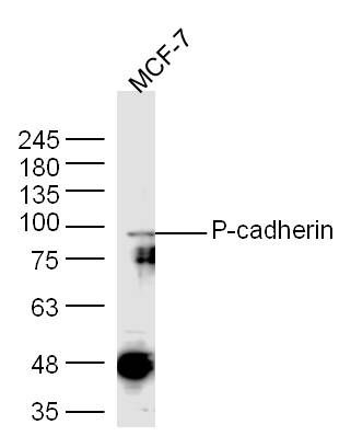
Sample:
MCF-7(Human) Cell Lysate at 30 ug
Primary: Anti-P-cadherin (SL1159R) at 1/300 dilution
Secondary: IRDye800CW Goat Anti-Rabbit IgG at 1/20000 dilution
Predicted band size: 80 kD
Observed band size: 90 kD
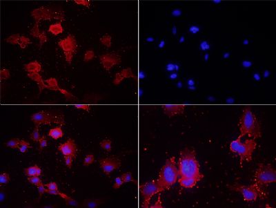
Tissue/cell: U251 cells;4% Paraformaldehyde-fixed;
Blocking buffer (normal goat serum,SLC0005) at 37℃ for 20 min;
Incubation: Anti-P-cadherin Polyclonal Antibody, Unconjugated(SL1159R) 1:200, overnight at 4°C; The secondary antibody was Goat Anti-Rabbit IgG, Cy3 conjugated(SL0295G-Cy3)used at 1:200 dilution for 40 minutes at 37°C. DAPI(5ug/ml,blue,SLC0033) was used to stain the cell nuclei
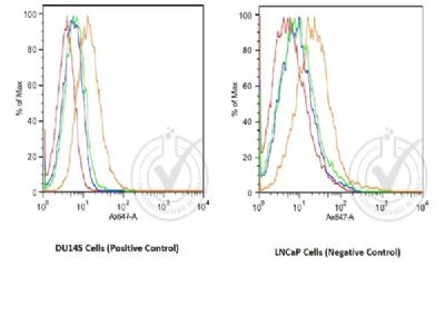
Image provided by Independent Validation (badge 029741). Histogram of human DU145 and human LNCaP cells stained with Rabbit Anti-P-cadherin Polyclonal Antibody (orange)(SL1159R at 1:100), isotype control antibody (green), secondary antibody only (blue) and unstained (red)
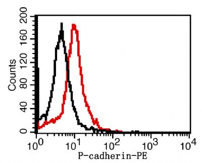
Overlay histogram showing Mouse SP2/0 stained with SL1159R-PE (red line). The cells were fixed with 1% paraformaldehyde (10 min). The cells were then incubated with the antibody(SL1159R-PE, 2ug/1x106cells) for 30 min at 22-25°C. Isotype control antibody (black line) was rabbit IgG (2ug/1x106cells) used under the same conditions. Acquisition of >5,000 events was performed.
|