Home > Product > Antibody > Rabbit Anti-presenilin 1 antibody
Presenilin-1 NTF subunit; AD 3; AD3; Ad3h; Alzheimer Disease 3; EC 3.4.23.; FAD; Homo Sapiens Clone CC44 Senilin 1; Presenilin 1 Alzheimer disease 3; Presenilin 1; Presenilin1; Protein S182; PS 1; PS-1; PS1; PSEN 1; PSEN1; PSN 1; PSN1; PSN1_HUMAN; PSNL 1;
Cat:
SL0024R
Species Reactivity:
Human,Mouse,Rat,(predicted: Dog,Pig,Cow,Rabbit,)
Immunogen:
KLH conjugated synthetic peptide derived from human Presenilin-1 NTF subunit:10-80/467
Format:
Liquid
Storage instructions:
Shipped at 4℃. Store at -20 °C for one year. Avoid repeated freeze/thaw cycles.
Concentration:
1mg/ml
Clonality:
Polyclonal
Isotype:
IgG
Applications:
WB=1:500-2000ELISA=1:5000-10000IHC-P=1:100-500IHC-F=1:100-500Flow-Cyt=1ug/testICC=1:100IF=1:100-500(Paraffin sections need to do antigen repair)not yet tested in other applications.optimal dilutions/concentrations should be determined by the end user.
Host:
Rabbit
Product Overview:
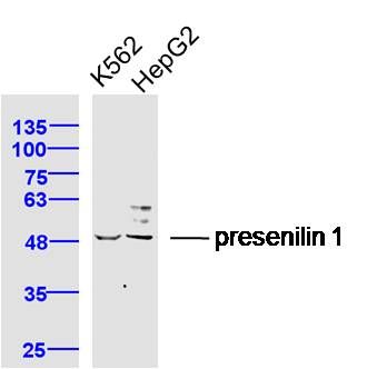
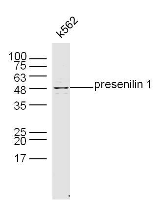
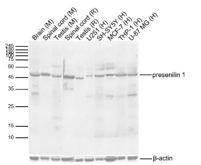
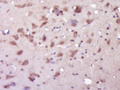
Sample: K562 Cell (Human) Lysate at 40 ugHepG2 Cell (Human) Lysate at 40 ugPrimary: Anti-presenilin 1 (SL0024R) at 1/300 dilutionSecondary: IRDye800CW Goat Anti-Rabbit IgG at 1/20000 dilutionPredicted band size: 34/52 kDObserved band size: 50 kDSample: K562 Cell Lysate at 40 ugPrimary: Anti- presenilin1 (SL0024R) at 1/300 dilutionSecondary: IRDye800CW Goat Anti-Rabbit IgG at 1/20000 dilutionPredicted band size: 34/52 kDObserved band size: 48 kDSample: Lane 1: Mouse Brain Lysates Lane 2: Mouse Spinal cord Lysates Lane 3: Mouse Testis Lysates Lane 4: Rat Spinal cord Lysates Lane 5: Rat Testis Lysates Lane 6: Human U251 cell Lysates Lane 7: Human SH-SY5Y cell Lysates Lane 8: Human MCF-7 cell Lysates Lane 9: Human THP-1 cell Lysates Lane 10: Human U-87 MG cell Lysates Primary: Anti-presenilin 1 (SL0024R) at 1/1000 dilutionSecondary: IRDye800CW Goat Anti-Rabbit IgG at 1/20000 dilutionPredicted band size: 34/52kDaObserved band size: 50kDaParaformaldehyde-fixed, paraffin embedded (Rat brain); Antigen retrieval by boiling in sodium citrate buffer (pH6.0) for 15min; Block endogenous peroxidase by 3% hydrogen peroxide for 20 minutes; Blocking buffer (normal goat serum) at 37°C for 30min; Antibody incubation with (presenilin 1) Polyclonal Antibody, Unconjugated (SL0024R) at 1:400 overnight at 4°C, followed by operating according to SP Kit(Rabbit) (sp-0023) instructions and DAB staining.SH-SY5Y cell; 4% Paraformaldehyde-fixed; Triton X-100 at room temperature for 20 min; Blocking buffer (normal goat serum, SLC0005) at 37°C for 20 min; Antibody incubation with (presenilin 1) polyclonal Antibody, Unconjugated (SL0024R) 1:100, 90 minutes at 37°C; followed by a conjugated Goat Anti-Rabbit IgG antibody at 37°C for 90 minutes, DAPI (blue, C02-04002) was used to stain the cell nuclei.Blank control: Raji. Primary Antibody (green line): Rabbit Anti-presenilin 1 antibody (SL0024R) Dilution: 1μg /10^6 cells; Isotype Control Antibody (orange line): Rabbit IgG . Secondary Antibody : Goat anti-rabbit IgG-PEDilution: 1μg /test. ProtocolThe cells were fixed with 4% PFA (10min at room temperature)and then permeabilized with PBST for 20 min at room temperature. The cells were then incubated in 5%BSA to block non-specific protein-protein interactions for 30 min at at room temperature .Cells stained with Primary Antibody for 30 min at room temperature. The secondary antibody used for 40 min at room temperature. Acquisition of 20,000 events was performed.
More
Unit:
Price: $
Product PDFs
Datasheet:


Alzheimer's disease (AD) patients with an inherited form of the disease carry mutations in the presenilin proteins (PSEN1; PSEN2) or in the amyloid precursor protein (APP). These disease-linked mutations result in increased production of the longer form of amyloid-beta (main component of amyloid deposits found in AD brains). Presenilins are postulated to regulate APP processing through their effects on gamma-secretase, an enzyme that cleaves APP. Also, it is thought that the presenilins are involved in the cleavage of the Notch receptor, such that they either directly regulate gamma-secretase activity or themselves are protease enzymes. Several alternatively spliced transcript variants encoding different isoforms have been identified for this gene, the full-length nature of only some have been determined. [provided by RefSeq, Aug 2008]

Function:
Probable catalytic subunit of the gamma-secretase complex, an endoprotease complex that catalyzes the intramembrane cleavage of integral membrane proteins such as Notch receptors and APP (beta-amyloid precursor protein). Requires the other members of the gamma-secretase complex to have a protease activity. May play a role in intracellular signaling and gene expression or in linking chromatin to the nuclear membrane. Stimulates cell-cell adhesion though its association with the E-cadherin/catenin complex. Under conditions of apoptosis or calcium influx, cleaves E-cadherin promoting the disassembly of the E-cadherin/catenin complex and increasing the pool of cytoplasmic beta-catenin, thus negatively regulating Wnt signaling. May also play a role in hematopoiesis.

Subunit:
Homodimer. Component of the gamma-secretase complex, a complex composed of a presenilin homodimer (PSEN1 or PSEN2), nicastrin (NCSTN), APH1 (APH1A or APH1B) and PEN2. Such minimal complex is sufficient for secretase activity. Other components which are associated with the complex include SLC25A64, SLC5A7, PHB and PSEN1 isoform 3. Predominantly heterodimer of a N-terminal (NTF) and a SLCterminal (CTF) endoproteolytical fragment. Associates with proteolytic processed SLCterminal fragments C83 and C99 of the amyloid precursor protein (APP). Associates with NOTCH1. Associates with cadherin/catenin adhesion complexes through direct binding to CDH1 or CDH2. Interaction with CDH1 stabilizes the complex and stimulates cell-cell aggregation. Interaction with CDH2 is essential for trafficking of CDH2 from the endoplasmic reticulum to the plasma membrane. Interacts with CTNND2, CTNNB1, HERPUD1, FLNA, FLNB, MTCH1, PKP4 and PARL. Interacts through its N-terminus with isoform 3 of GFAP. Interacts with DOCK3.

Subcellular Location:
Endoplasmic reticulum membrane; Multi-pass membrane protein. Golgi apparatus membrane; Multi-pass membrane protein. Cell surface. Note=Bound to NOTCH1 also at the cell surface. Colocalizes with CDH1/2 at sites of cell-cell contact. Colocalizes with CTNNB1 in the endoplasmic reticulum and the proximity of the plasma membrane. Also present in azurophil granules of neutrophils.

Tissue Specificity:
Expressed in a wide range of tissues including various regions of the brain, liver, spleen and lymph nodes.

Post-translational modifications:
Heterogeneous proteolytic processing generates N-terminal (NTF) and SLCterminal (CTF) fragments of approximately 35 and 20 kDa, respectively. During apoptosis, the SLCterminal fragment (CTF) is further cleaved by caspase-3 to produce the fragment, PS1-CTF12.
After endoproteolysis, the SLCterminal fragment (CTF) is phosphorylated on serine residues by PKA and/or PKC. Phosphorylation on Ser-346 inhibits endoproteolysis.

DISEASE:
Defects in PSEN1 are a cause of Alzheimer disease type 3 (AD3) [MIM:607822]. AD3 is a familial early-onset form of Alzheimer disease. Alzheimer disease is a neurodegenerative disorder characterized by progressive dementia, loss of cognitive abilities, and deposition of fibrillar amyloid proteins as intraneuronal neurofibrillary tangles, extracellular amyloid plaques and vascular amyloid deposits. The major constituent of these plaques is the neurotoxic amyloid-beta-APP 40-42 peptide (s), derived proteolytically from the transmembrane precursor protein APP by sequential secretase processing. The cytotoxic SLCterminal fragments (CTFs) and the caspase-cleaved products such as C31 derived from APP, are also implicated in neuronal death.
Defects in PSEN1 are a cause of frontotemporal dementia (FTD) [MIM:600274].

Similarity:
Belongs to the peptidase A22A family.

SWISS:
P49768

Gene ID:
5663

Database links:

Entrez Gene: 5663 Human

Entrez Gene: 19164 Mouse

Entrez Gene: 29192 Rat

Omim: 104311 Human

SwissProt: P49768 Human

SwissProt: P49769 Mouse

SwissProt: P97887 Rat

Unigene: 3260 Human

Unigene: 998 Mouse

Unigene: 4488 Rat


此抗体识别分子量为45-50kDa早老素蛋白-1。PS-1主要在神经细胞中表达,早老蛋白集中于体细胞和树突状细胞中。相反,再早发家族AD(FAD)中和散发AD病人中,PS1免疫反应出现在老年斑和神经纤维缠结的神经炎中。 Picture

Sample:
K562 Cell (Human) Lysate at 40 ug
HepG2 Cell (Human) Lysate at 40 ug
Primary: Anti-presenilin 1 (SL0024R) at 1/300 dilution
Secondary: IRDye800CW Goat Anti-Rabbit IgG at 1/20000 dilution
Predicted band size: 34/52 kD
Observed band size: 50 kD
Sample: K562 Cell Lysate at 40 ug
Primary: Anti- presenilin1 (SL0024R) at 1/300 dilution
Secondary: IRDye800CW Goat Anti-Rabbit IgG at 1/20000 dilution
Predicted band size: 34/52 kD
Observed band size: 48 kD
Sample:
Lane 1: Mouse Brain Lysates
Lane 2: Mouse Spinal cord Lysates
Lane 3: Mouse Testis Lysates
Lane 4: Rat Spinal cord Lysates
Lane 5: Rat Testis Lysates
Lane 6: Human U251 cell Lysates
Lane 7: Human SH-SY5Y cell Lysates
Lane 8: Human MCF-7 cell Lysates
Lane 9: Human THP-1 cell Lysates
Lane 10: Human U-87 MG cell Lysates
Primary: Anti-presenilin 1 (SL0024R) at 1/1000 dilution
Secondary: IRDye800CW Goat Anti-Rabbit IgG at 1/20000 dilution
Predicted band size: 34/52kDa
Observed band size: 50kDa
Paraformaldehyde-fixed, paraffin embedded (Rat brain); Antigen retrieval by boiling in sodium citrate buffer (pH6.0) for 15min; Block endogenous peroxidase by 3% hydrogen peroxide for 20 minutes; Blocking buffer (normal goat serum) at 37°C for 30min; Antibody incubation with (presenilin 1) Polyclonal Antibody, Unconjugated (SL0024R) at 1:400 overnight at 4°C, followed by operating according to SP Kit(Rabbit) (sp-0023) instructions and DAB staining.
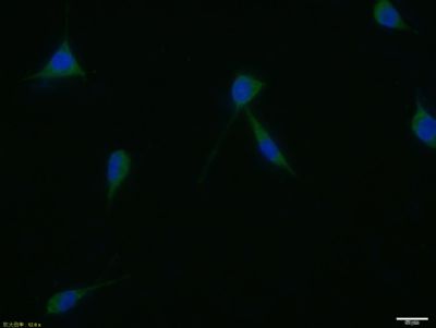
SH-SY5Y cell; 4% Paraformaldehyde-fixed; Triton X-100 at room temperature for 20 min; Blocking buffer (normal goat serum, SLC0005) at 37°C for 20 min; Antibody incubation with (presenilin 1) polyclonal Antibody, Unconjugated (SL0024R) 1:100, 90 minutes at 37°C; followed by a conjugated Goat Anti-Rabbit IgG antibody at 37°C for 90 minutes, DAPI (blue, C02-04002) was used to stain the cell nuclei.
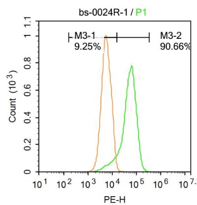
Blank control: Raji.
Primary Antibody (green line): Rabbit Anti-presenilin 1 antibody (SL0024R)
Dilution: 1μg /10^6 cells;
Isotype Control Antibody (orange line): Rabbit IgG .
Secondary Antibody : Goat anti-rabbit IgG-PE
Dilution: 1μg /test.
Protocol
The cells were fixed with 4% PFA (10min at room temperature)and then permeabilized with PBST for 20 min at room temperature. The cells were then incubated in 5%BSA to block non-specific protein-protein interactions for 30 min at at room temperature .Cells stained with Primary Antibody for 30 min at room temperature. The secondary antibody used for 40 min at room temperature. Acquisition of 20,000 events was performed.
Product Feedback Wall
Message :
Your Email :
Copyright © 2007-2018 Sunlong Medical All Rights Reserved.