Home > Product > Antibody > Rabbit Anti-TGF beta Receptor I antibody
TGF-beta receptor type-1; TGFBR1; TGF Beta R1; AAT 5; AAT5; Activin A receptor type II like kinase 53kDa; Activin receptor like kinase 5; ACVRLK 4; ACVRLK4; ALK 5; ALK5; Serine/threonine protein kinase receptor R4; SKR 4; SKR4; TbetaR I; TGF beta receptor
Cat:
SL0638R
Species Reactivity:
Human,Mouse,Rat,(predicted: Cow,)
Immunogen:
KLH conjugated synthetic peptide derived from human TGF beta Receptor I :301-400/501<Extracellular>
Format:
Liquid
Storage instructions:
Shipped at 4℃. Store at -20 °C for one year. Avoid repeated freeze/thaw cycles.
Concentration:
1mg/ml
Clonality:
Polyclonal
Isotype:
IgG
Applications:
WB=1:500-2000ELISA=1:5000-10000IHC-P=1:100-500IHC-F=1:100-500Flow-Cyt=1ug/TestIF=1:100-500(Paraffin sections need to do antigen repair)not yet tested in other applications.optimal dilutions/concentrations should be determined by the end user.
Host:
Rabbit
Product Overview:
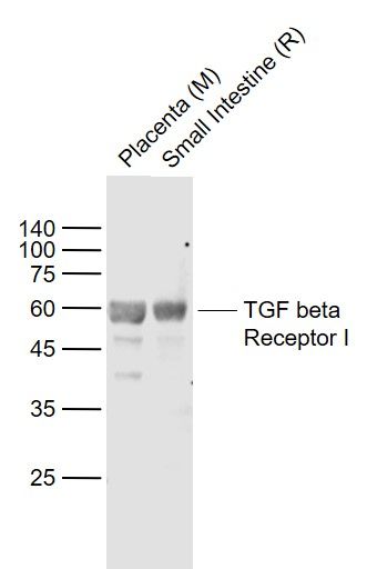
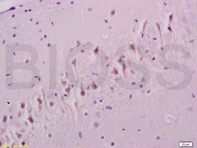
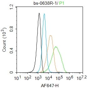
Sample: Lane 1: Placenta (Mouse) Lysate at 40 ug Lane 2: Small intestine (Rat) Lysate at 40 ug Primary: Anti- TGF beta Receptor I (SL0638R) at 1/1000 dilution Secondary: IRDye800CW Goat Anti-Rabbit IgG at 1/20000 dilution Predicted band size: 56 kD Observed band size: 58 kDTissue/cell: rat brain tissue; 4% Paraformaldehyde-fixed and paraffin-embedded; Antigen retrieval: citrate buffer ( 0.01M, pH 6.0 ), Boiling bathing for 15min; Block endogenous peroxidase by 3% Hydrogen peroxide for 30min; Blocking buffer (normal goat serum,SLC0005) at 37∩ for 20 min; Incubation: Anti-TGF Beta R1/TGFBR1 Polyclonal Antibody, Unconjugated(SL0638R) 1:200, overnight at 4∑C, followed by conjugation to the secondary antibody(SP-0023) and DAB(SLC0010) stainingBlank control:U937. Primary Antibody (green line): Rabbit Anti-TGF beta Receptor I antibody (SL0638R) Dilution: 1μg /10^6 cells; Isotype Control Antibody (orange line): Rabbit IgG . Secondary Antibody : Goat anti-rabbit IgG-AF647Dilution: 1μg /test. ProtocolThe cells were incubated in 5%BSA to block non-specific protein-protein interactions for 30 min at room temperature .Cells stained with Primary Antibody for 30 min at room temperature. The secondary antibody used for 40 min at room temperature. Acquisition of 20,000 events was performed.
More
Unit:
Price: $
Product PDFs
Datasheet:


The protein encoded by this gene forms a heteromeric complex with type II TGF-beta receptors when bound to TGF-beta, transducing the TGF-beta signal from the cell surface to the cytoplasm. The encoded protein is a serine/threonine protein kinase. Mutations in this gene have been associated with Loeys-Dietz aortic aneurysm syndrome (LDAS). Multiple transcript variants encoding different isoforms have been found for this gene. [provided by RefSeq, Aug 2008]

Function:
On ligand binding, forms a receptor complex consisting of two type II and two type I transmembrane serine/threonine kinases. Type II receptors phosphorylate and activate type I receptors which autophosphorylate, then bind and activate SMAD transcriptional regulators. Receptor for TGF-beta.

Subunit:
Homodimer; in the endoplasmic reticulum but also at the cell membrane. Heterohexamer; TGFB1, TGFB2 and TGFB3 homodimeric ligands assemble a functional receptor composed of two TGFBR1 and TGFBR2 heterodimers to form a ligand-receptor heterohexamer. The respective affinity of TGBRB1 and TGFBR2 for the ligands may modulate the kinetics of assembly of the receptor and may explain the different biological activities of TGFB1, TGFB2 and TGFB3. Interacts with CD109; inhibits TGF-beta receptor activation in keratinocytes. Interacts with RBPMS. Interacts (unphosphorylated) with FKBP1A; prevents TGFBR1 phosphorylation by TGFBR2 and stabilizes it in the inactive conformation. Interacts with SMAD2, SMAD3 and ZFYVE9; ZFYVE9 recruits SMAD2 and SMAD3 to the TGF-beta receptor. Interacts with TRAF6 and MAP3K7; induces MAP3K7 activation by TRAF6. Interacts with PARD6A; involved in TGF-beta induced epithelial to mesenchymal transition. Interacts with SMAD7, NEDD4L, SMURF1 and SMURF2; SMAD7 recruits NEDD4L, SMURF1 and SMURF2 to the TGF-beta receptor.

Subcellular Location:
Cell membrane; Single-pass type I membrane protein. Cell junction, tight junction.

Tissue Specificity:
Found in all tissues examined, most abundant in placenta and least abundant in brain and heart.

Post-translational modifications:
Phosphorylated at basal levels in the absence of ligand binding. Activated by multiple phosphorylation, mainly in the GS region.

Similarity:
Belongs to the protein kinase superfamily. TKL Ser/Thr protein kinase family. TGFB receptor subfamily.
Contains 1 GS domain.
Contains 1 protein kinase domain.

SWISS:
P36897

Gene ID:
7046

Database links:

Entrez Gene: 7046 Human

Entrez Gene: 21812 Mouse

Entrez Gene: 29591 Rat

Entrez Gene: 282382 Cow

Omim: 190181 Human

SwissProt: O46136 Cow

SwissProt: P36897 Human

SwissProt: Q64729 Mouse

SwissProt: P80204 Rat

Unigene: 494622 Human

Unigene: 197552 Mouse

Unigene: 4882 Rat


细胞膜受体(Membrane Receptors)TGF-βRⅠ Picture

Sample:
Lane 1: Placenta (Mouse) Lysate at 40 ug
Lane 2: Small intestine (Rat) Lysate at 40 ug
Primary: Anti- TGF beta Receptor I (SL0638R) at 1/1000 dilution
Secondary: IRDye800CW Goat Anti-Rabbit IgG at 1/20000 dilution
Predicted band size: 56 kD
Observed band size: 58 kD
Tissue/cell: rat brain tissue; 4% Paraformaldehyde-fixed and paraffin-embedded;
Antigen retrieval: citrate buffer ( 0.01M, pH 6.0 ), Boiling bathing for 15min; Block endogenous peroxidase by 3% Hydrogen peroxide for 30min; Blocking buffer (normal goat serum,SLC0005) at 37∩ for 20 min;
Incubation: Anti-TGF Beta R1/TGFBR1 Polyclonal Antibody, Unconjugated(SL0638R) 1:200, overnight at 4∑C, followed by conjugation to the secondary antibody(SP-0023) and DAB(SLC0010) staining
Blank control:U937.
Primary Antibody (green line): Rabbit Anti-TGF beta Receptor I antibody (SL0638R)
Dilution: 1μg /10^6 cells;
Isotype Control Antibody (orange line): Rabbit IgG .
Secondary Antibody : Goat anti-rabbit IgG-AF647
Dilution: 1μg /test.
Protocol
The cells were incubated in 5%BSA to block non-specific protein-protein interactions for 30 min at room temperature .Cells stained with Primary Antibody for 30 min at room temperature. The secondary antibody used for 40 min at room temperature. Acquisition of 20,000 events was performed.
Product Feedback Wall
Message :
Your Email :
Copyright © 2007-2018 Sunlong Medical All Rights Reserved.