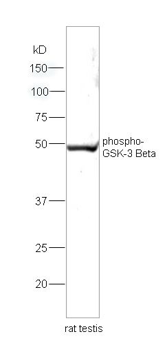
[IF=6.222] Wang, Lili. et al. Progranulin improves neural development via the PI3K/Akt/GSK-3β pathway in the cerebellum of a VPA-induced rat model of ASD. Transl Psychiat. 2022 Mar;12(1):1-14 WB ; Rat.
[IF=5.279] Shuhua Tian. et al. Sulforaphane Regulates Glucose and Lipid Metabolisms in Obese Mice by Restraining JNK and Activating Insulin and FGF21 Signal Pathways. J Agr Food Chem. 2021;XXXX(XXX):XXX-XXX WB ; Mouse.
[IF=6.023] Ling Xie. et al. Suppression of GOLM1 by EGCG through HGF/HGFR/AKT/GSK-3β/β-catenin/c-Myc signaling pathway inhibits cell migration of MDA-MB-231. Food Chem Toxicol. 2021 Nov;157:112574 WB ; human.
[IF=5.25] Junying Lan. et al. Abnormal spatiotemporal expression pattern of progranulin and neurodevelopment impairment in VPA-induced ASD rat model. Neuropharmacology. 2021 Sep;196:108689 WB ; Rat.
[IF=7.971] He J et al. SLC34A2 Simultaneously Promotes Papillary Thyroid Carcinoma Growth and Invasion Through Distinct Mechanisms. Oncogene. 2020 Mar;39(13):2658-2675. WB ; human.
[IF=5.5] Wang Y et al. High Concentration of Aspirin Induces Apoptosis in Rat Tendon Stem Cells via Inhibition of the Wnt/β-Catenin Pathway. (2018) Cell Physiol Biochem;50(6):2046-2059. WB ; Rat.
[IF=0.23] Li, Zhao, et al. "The aqueous extract of Curcuma wenyujin rescues learning and memory deficits through PI3k/Akt/GSK-3β pathway in Aβ-induced AD mice." Biomedical Research 28.17 (2017): 7438-7442. WB ; Mouse.
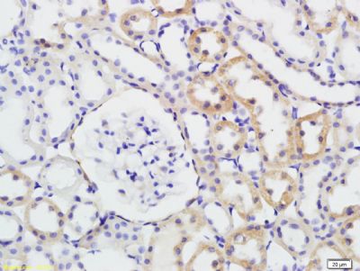
[IF=4.803] Liqin An. et al. Bone Morphogenetic Protein 4 (BMP4) promotes hepatic glycogen accumulation and reduces glucose level in hepatocytes through mTORC2 signaling pathway. Genes Dis. 2020 Nov;: WB,IHC ; Mouse.
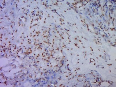
[IF=2.396] Ren et al. GSK-3β inhibits autophagy and enhances radiosensitivity in non-small cell lung cancer. (2018) Diagn.Pathol. 13:33 IHC ; Human.
[IF=2.833] Peiqi Zhu. et al. Panax notoginseng saponins promote endothelial progenitor cell angiogenesis via the Wnt/β-catenin pathway. Bmc Complem Altern M. 2021 Dec;21(1):1-11 WB ; Dog.
[IF=1.918] Qingxin Fan. et al. Ginsenoside Rb1 Facilitates Browning by Repressing Wnt/β-Catenin Signaling in 3T3-L1 Adipocytes. Med Sci Monitor. 2021; 27: e928619-1–e928619-10 WB ;
[IF=6.17] Tong Xu. et al. Lithium chloride represses abdominal aortic aneurysm via regulating GSK3β/SIRT1/NF-κB signaling pathway. Free Radical Bio Med. 2021 Apr;166:1 WB,IHC ; Rat.
[IF=5.52] Mikami, Norihisa, et al. "Calcitonin gene-related peptide and cyclic adenosine 5'-monophosphate/protein kinase A pathway promote IL-9 production in Th9 differentiation process. 2013 Apr 15;190(8):4046-55.
[IF=2.884] Sun L et al. MiR-26a promotes fracture healing of nonunion rats possibly by targeting SOSTDC1 and further activating Wnt/β-catenin signaling pathway. Mol Cell Biochem. 2019 Jul 16. WB ; Rat.
[IF=2.396] Ren J et al. GSK-3β inhibits autophagy and enhances radiosensitivity in non-small cell lung cancer.Diagn Pathol. 2018 May 24;13(1):33. IHSLCP ; Human.
[IF=2.766] Zhang et al. Over-Expressed Twist Associates with Markers of Epithelial Mesenchymal Transition and Predicts Poor Prognosis in Breast Cancers via ERK and Akt Activation. (2015) PLoS.One. 10:e0135851 WB ; Human.