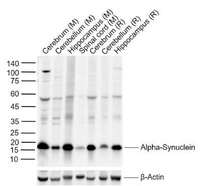
Sample:
Lane 1: Mouse Cerebrum tissue lysates
Lane 2: Mouse Cerebellum tissue lysates
Lane 3: Mouse Hippocampus tissue lysates
Lane 4: Mouse Spinal cord tissue lysates
Lane 5: Rat Cerebrum tissue lysates
Lane 6: Rat Cerebellum tissue lysates
Lane 7: Rat Hippocampus tissue lysates
Primary: Anti-Alpha-Synuclein (SL34113R) at 1/1000 dilution
Secondary: IRDye800CW Goat Anti-Rabbit IgG at 1/20000 dilution
Predicted band size: 14 kDa
Observed band size: 16 kDa
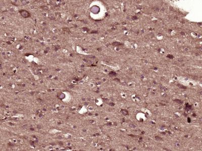
Paraformaldehyde-fixed, paraffin embedded (Human brain glioma); Antigen retrieval by boiling in sodium citrate buffer (pH6.0) for 15min; Block endogenous peroxidase by 3% hydrogen peroxide for 20 minutes; Blocking buffer (normal goat serum) at 37°C for 30min; Antibody incubation with (alpha-Synuclein) Polyclonal Antibody, Unconjugated (SL0009R) at 1:400 overnight at 4°C, followed by operating according to SP Kit(Rabbit) (sp-0023) instructionsand DAB staining.
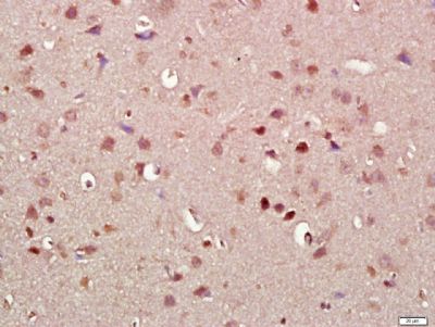
Paraformaldehyde-fixed, paraffin embedded (Rat brain); Antigen retrieval by boiling in sodium citrate buffer (pH6.0) for 15min; Block endogenous peroxidase by 3% hydrogen peroxide for 20 minutes; Blocking buffer (normal goat serum) at 37°C for 30min; Antibody incubation with (alpha-Synuclein) Polyclonal Antibody, Unconjugated (SL0009R) at 1:500 overnight at 4°C, followed by a conjugated secondary (sp-0023) for 20 minutes and DAB staining.
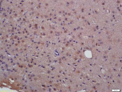
Paraformaldehyde-fixed, paraffin embedded (rat brain tissue); Antigen retrieval by boiling in sodium citrate buffer (pH6.0) for 15min; Block endogenous peroxidase by 3% hydrogen peroxide for 20 minutes; Blocking buffer (normal goat serum) at 37°C for 30min; Antibody incubation with (alpha-Synuclein) Polyclonal Antibody, Unconjugated (SL0009R) at 1:400 overnight at 4°C, followed by a conjugated secondary (sp-0023) for 20 minutes and DAB staining.
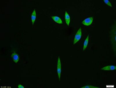
SH-SY5Y cell; 4% Paraformaldehyde-fixed; Triton X-100 at room temperature for 20 min; Blocking buffer (normal goat serum, SLC0005) at 37°C for 20 min; Antibody incubation with (alpha-Synuclein) polyclonal Antibody, Unconjugated (SL0009R) 1:100, 90 minutes at 37°C; followed by a conjugated Goat Anti-Rabbit IgG antibody at 37°C for 90 minutes, DAPI (blue, C02-04002) was used to stain the cell nuclei.
|