Home > Product > Antibody > Rabbit Anti-HNF1A antibody
HNF1A; TCF1; HNF1 alpha; HNF-1 Alpha; Hepatocyte nuclear factor 1 Alpha; Albumin proximal factor; Hepatic nuclear factor 1 alpha; Hepatic nuclear factor 1; Hepatic transcription factor 1 alpha; Hepatic transcription factor 1; HNF 1; HNF 1A; HNF1A; Interfe
Cat:
SL1405R
Species Reactivity:
Human,Mouse,Rat,(predicted: Chicken,Dog,Pig,Cow,Zebrafish,Sheep,)
Immunogen:
KLH conjugated synthetic peptide derived from human HNF1:201-350/628
Format:
Liquid
Storage instructions:
Shipped at 4℃. Store at -20 °C for one year. Avoid repeated freeze/thaw cycles.
Concentration:
1mg/ml
Clonality:
Polyclonal
Isotype:
IgG
Applications:
WB=1:500-2000ELISA=1:5000-10000IHC-P=1:100-500Flow-Cyt=3ug/testICC=1:100(Paraffin sections need to do antigen repair)not yet tested in other applications.optimal dilutions/concentrations should be determined by the end user.
Host:
Rabbit
Product Overview:
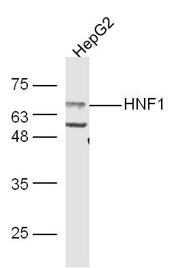
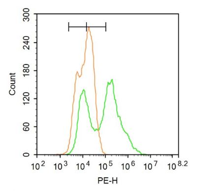
Sample: HepG2 Cell (Human) Lysate at 30 ugPrimary: Anti-HNF1 (Bs-1405R) at 1/300 dilutionSecondary: IRDye800CW Goat Anti-Rabbit IgG at 1/20000 dilutionPredicted band size: 67 kDObserved band size: 67 kD Paraformaldehyde-fixed, paraffin embedded (Mouse liver); Antigen retrieval by boiling in sodium citrate buffer (pH6.0) for 15min; Block endogenous peroxidase by 3% hydrogen peroxide for 20 minutes; Blocking buffer (normal goat serum) at 37°C for 30min; Antibody incubation with (HNF1A) Polyclonal Antibody, Unconjugated (SL1405R) at 1:400 overnight at 4°C, followed by operating according to SP Kit(Rabbit) (sp-0023) instructionsand DAB staining. Paraformaldehyde-fixed, paraffin embedded (Rat liver); Antigen retrieval by boiling in sodium citrate buffer (pH6.0) for 15min; Block endogenous peroxidase by 3% hydrogen peroxide for 20 minutes; Blocking buffer (normal goat serum) at 37°C for 30min; Antibody incubation with (HNF1A) Polyclonal Antibody, Unconjugated (SL1405R) at 1:400 overnight at 4°C, followed by operating according to SP Kit(Rabbit) (sp-0023) instructionsand DAB staining.HepG2 cell; 4% Paraformaldehyde-fixed; Triton X-100 at room temperature for 20 min; Blocking buffer (normal goat serum, SLC0005) at 37°C for 20 min; Antibody incubation with (HNF1A) polyclonal Antibody, Unconjugated (SL1405R) 1:100, 90 minutes at 37°C; followed by a conjugated Goat Anti-Rabbit IgG antibody at 37°C for 90 minutes, DAPI (blue, C02-04002) was used to stain the cell nuclei.Blank control:A549. Primary Antibody (green line): Rabbit Anti-HNF1A antibody (SL1405R) Dilution: 1μg /10^6 cells; Isotype Control Antibody (orange line): Rabbit IgG . Secondary Antibody : Goat anti-rabbit IgG-PEDilution: 3μg /test. ProtocolThe cells were fixed with 4% PFA (10min at room temperature)and then permeabilized with 90% ice-cold methanol for 20 min at-20℃. The cells were then incubated in 5% BSA to block non-specific protein-protein interactions for 30 min at at room temperature .Cells stained with Primary Antibody for 30 min at room temperature. The secondary antibody used for 40 min at room temperature. Acquisition of 20,000 events was performed.
More
Unit:
Price: $
Product PDFs
Datasheet:


IRF1 encodes interferon regulatory factor 1, a member of the interferon regulatory transcription factor (IRF) family. IRF1 serves as an activator of interferons alpha and beta transcription, and in mouse it has been shown to be required for double-stranded RNA induction of these genes. IRF1 also functions as a transcription activator of genes induced by interferons alpha, beta, and gamma. Further, IRF1 has been shown to play roles in regulating apoptosis and tumor-suppressoion.

Function:
Transcriptional activator that regulates the tissue specific expression of multiple genes, especially in pancreatic islet cells and in liver. Required for the expression of several liver specific genes. Binds to the inverted palindrome 5'-GTTAATNATTAASLC3'.

Subunit:
Binds DNA as a dimer. Interacts with PCBD1. Heterotetramer with PCBD1; formed by a dimer of dimers.

Subcellular Location:
Nucleus. [ALTERNATIVE PRODUCTS] Event=Alternative splicing; Named isoforms=3; Name=A; IsoId=P20823-1; Sequence=Displayed; Name=B; IsoId=P20823-2; Sequence=VSP_002250, VSP_002251; Name=C; IsoId=P20823-3; Sequence=VSP_002252, VSP_002253.

Tissue Specificity:
Liver.

DISEASE:
Hepatic adenomas familial (HEPAF) [MIM:142330]: Rare benign liver tumors of presumable epithelial origin that develop in an otherwise normal liver. Hepatic adenomas may be single or multiple. They consist of sheets of well-differentiated hepatocytes that contain fat and glycogen and can produce bile. Bile ducts or portal areas are absent. Kupffer cells, if present, are reduced in number and are non-functional. Conditions associated with adenomas are insulin-dependent diabetes mellitus and glycogen storage diseases (types 1 and 3). Note=The disease is caused by mutations affecting the gene represented in this entry. Bi-allelic inactivation of HNF1A, whether sporadic or associated with MODY3, may be an early step in the developmant of some hepatocellular carcinomas.
Maturity-onset diabetes of the young 3 (MODY3) [MIM:600496]: A form of diabetes that is characterized by an autosomal dominant mode of inheritance, onset in childhood or early adulthood (usually before 25 years of age), a primary defect in insulin secretion and frequent insulin-independence at the beginning of the disease. Note=The disease is caused by mutations affecting the gene represented in this entry.
Diabetes mellitus, insulin-dependent, 20 (IDDM20) [MIM:612520]: A multifactorial disorder of glucose homeostasis that is characterized by susceptibility to ketoacidosis in the absence of insulin therapy. Clinical fetaures are polydipsia, polyphagia and polyuria which result from hyperglycemia-induced osmotic diuresis and secondary thirst. These derangements result in long-term complications that affect the eyes, kidneys, nerves, and blood vessels. Note=Disease susceptibility is associated with variations affecting the gene represented in this entry.

Similarity:
Belongs to the HNF1 homeobox family.
Contains 1 homeobox DNA-binding domain.

SWISS:
P15257

Gene ID:
6927

Database links:

Entrez Gene: 6927 Human

Entrez Gene: 21405 Mouse

Entrez Gene: 574067 Pig

Entrez Gene: 24817 Rat

Omim: 142410 Human

SwissProt: P20823 Human

SwissProt: P22361 Mouse

SwissProt: P15257 Rat

Unigene: 654455 Human

Unigene: 332607 Mouse

Unigene: 9660 Rat


转录调节因子(Transcriptin Regulators) Picture

Sample:
HepG2 Cell (Human) Lysate at 30 ug
Primary: Anti-HNF1 (Bs-1405R) at 1/300 dilution
Secondary: IRDye800CW Goat Anti-Rabbit IgG at 1/20000 dilution
Predicted band size: 67 kD
Observed band size: 67 kD
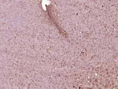
Paraformaldehyde-fixed, paraffin embedded (Mouse liver); Antigen retrieval by boiling in sodium citrate buffer (pH6.0) for 15min; Block endogenous peroxidase by 3% hydrogen peroxide for 20 minutes; Blocking buffer (normal goat serum) at 37°C for 30min; Antibody incubation with (HNF1A) Polyclonal Antibody, Unconjugated (SL1405R) at 1:400 overnight at 4°C, followed by operating according to SP Kit(Rabbit) (sp-0023) instructionsand DAB staining.
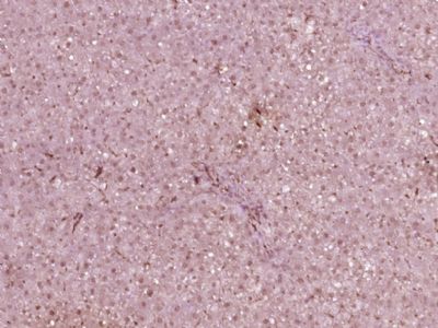
Paraformaldehyde-fixed, paraffin embedded (Rat liver); Antigen retrieval by boiling in sodium citrate buffer (pH6.0) for 15min; Block endogenous peroxidase by 3% hydrogen peroxide for 20 minutes; Blocking buffer (normal goat serum) at 37°C for 30min; Antibody incubation with (HNF1A) Polyclonal Antibody, Unconjugated (SL1405R) at 1:400 overnight at 4°C, followed by operating according to SP Kit(Rabbit) (sp-0023) instructionsand DAB staining.
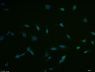
HepG2 cell; 4% Paraformaldehyde-fixed; Triton X-100 at room temperature for 20 min; Blocking buffer (normal goat serum, SLC0005) at 37°C for 20 min; Antibody incubation with (HNF1A) polyclonal Antibody, Unconjugated (SL1405R) 1:100, 90 minutes at 37°C; followed by a conjugated Goat Anti-Rabbit IgG antibody at 37°C for 90 minutes, DAPI (blue, C02-04002) was used to stain the cell nuclei.
Blank control:A549.
Primary Antibody (green line): Rabbit Anti-HNF1A antibody (SL1405R)
Dilution: 1μg /10^6 cells;
Isotype Control Antibody (orange line): Rabbit IgG .
Secondary Antibody : Goat anti-rabbit IgG-PE
Dilution: 3μg /test.
Protocol
The cells were fixed with 4% PFA (10min at room temperature)and then permeabilized with 90% ice-cold methanol for 20 min at-20℃. The cells were then incubated in 5% BSA to block non-specific protein-protein interactions for 30 min at at room temperature .Cells stained with Primary Antibody for 30 min at room temperature. The secondary antibody used for 40 min at room temperature. Acquisition of 20,000 events was performed.
Product Feedback Wall
Message :
Your Email :
Copyright © 2007-2018 Sunlong Medical All Rights Reserved.