Histone acetylation and deacetylation, catalyzed by multisubunit complexes, play a key role in the regulation of eukaryotic gene expression. The protein encoded by this gene belongs to the histone deacetylase/acuc/apha family and is a component of the histone deacetylase complex. It also interacts with retinoblastoma tumor-suppressor protein and this complex is a key element in the control of cell proliferation and differentiation. Together with metastasis-associated protein-2, it deacetylates p53 and modulates its effect on cell growth and apoptosis. [provided by RefSeq, Jul 2008]
Function:
Responsible for the deacetylation of lysine residues on the N-terminal part of the core histones (H2A, H2B, H3 and H4). Histone deacetylation gives a tag for epigenetic repression and plays an important role in transcriptional regulation, cell cycle progression and developmental events. Histone deacetylases act via the formation of large multiprotein complexes. Deacetylates SP proteins, SP1 and SP3, and regulates their function. Component of the BRG1-RB1-HDAC1 complex, which negatively regulates the CREST-mediated transcription in resting neurons. Upon calcium stimulation, HDAC1 is released from the complex and CREBBP is recruited, which facilitates transcriptional activation. Deacetylates TSHZ3 and regulates its transcriptional repressor activity. Deacetylates 'Lys-310' in RELA and thereby inhibits the transcriptional activity of NF-kappa-B. Component a RCOR/GFI/KDM1A/HDAC complex that suppresses, via histone deacetylase (HDAC) recruitment, a number of genes implicated in multilineage blood cell development.
Subcellular Location:
Nucleus.
Tissue Specificity:
Ubiquitous, with higher levels in heart, pancreas and testis, and lower levels in kidney and brain.
Post-translational modifications:
Sumoylated on Lys-444 and Lys-476; which promotes enzymatic activity. Desumoylated by SENP1.
Phosphorylation on Ser-421 and Ser-423 promotes enzymatic activity and interactions with NuRD and SIN3 complexes. Phosphorylated by CDK5.
Ubiquitinated by CHFR, leading to its degradation by the proteasome. Ubiquitinated by KCTD11, leading to proteasomal degradation.
Similarity:
Belongs to the histone deacetylase family. HD type 1 subfamily.
SWISS:
Q13547
Gene ID:
3065
Database links:
Entrez Gene: 3065 Human
Entrez Gene: 433759 Mouse
Entrez Gene: 297893 Rat
Entrez Gene: 404126 Cow
Omim: 601241 Human
SwissProt: Q32PJ8 Cow
SwissProt: Q13547 Human
SwissProt: O09106 Mouse
SwissProt: Q4QQW4 Rat
Unigene: 88556 Human
Unigene: 202504 Mouse
Unigene: 391033 Mouse
Unigene: 1863 Rat
组蛋白去乙酰化酶(HDACs)是一组在细胞染色质水平、通过诱导组蛋白去乙酰化来调控包括染色质重组、转录活化或抑制、细胞周期、细胞分化及细胞凋亡等一系列生物学效应的酶,特别是与细胞活化后的基因转录表达调控有关。
| Picture |
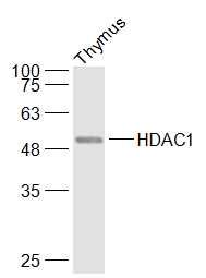
Sample:
Thymus (Mouse) Lysate at 40 ug
Primary: Anti-HDAC1 (SL1414R) at 1/1000 dilution
Secondary: IRDye800CW Goat Anti-Rabbit IgG at 1/20000 dilution
Predicted band size: 53 kD
Observed band size: 53 kD
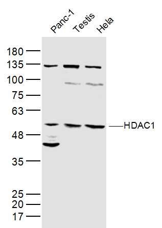
Sample:
Panc-1(Human) Cell Lysate at 40 ug
Testis (Mouse) Lysate at 40 ug
Hela(Human) Lysate at 40 ug
Primary: Anti-HDAC1 (SL1414R) at 1/300 dilution
Secondary: IRDye800CW Goat Anti-Rabbit IgG at 1/20000 dilution
Predicted band size: 53 kD
Observed band size: 53 kD
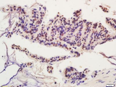
Tissue/cell: human colon carcinoma; 4% Paraformaldehyde-fixed and paraffin-embedded;
Antigen retrieval: citrate buffer ( 0.01M, pH 6.0 ), Boiling bathing for 15min; Block endogenous peroxidase by 3% Hydrogen peroxide for 30min; Blocking buffer (normal goat serum,SLC0005) at 37℃ for 20 min;
Incubation: Anti-HDAC1 Polyclonal Antibody, Unconjugated(SL1414R) 1:200, overnight at 4°C, followed by conjugation to the secondary antibody(SP-0023) and DAB(SLC0010) staining
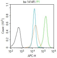
Blank control (Black line):Molt4 (Black).
Primary Antibody (green line): Rabbit Anti-HDAC1 antibody (SL1414R)
Dilution: 1μg /10^6 cells;
Isotype Control Antibody (orange line): Rabbit IgG .
Secondary Antibody (white blue line): Goat anti-rabbit IgG-AF647
Dilution: 1μg /test.
Protocol
The cells were fixed with 4% PFA (10min at room temperature)and then permeabilized with 90% ice-cold methanol for 20 min at room temperature. The cells were then incubated in 5%BSA to block non-specific protein-protein interactions for 30 min at room temperature .Cells stained with Primary Antibody for 30 min at room temperature. The secondary antibody used for 40 min at room temperature. Acquisition of 20,000 events was performed.
|
|
|