Home > Product > Antibody > Rabbit Anti-XBP1 antibody
X box-binding protein-1; XBP-1; XBP2; Tax responsive element binding protein 5; TREB5; X box binding protein 2; XBP 1; XBP2; XBP1_HUMAN; X-box-binding protein 1, cytoplasmic form; X-box-binding protein 1, luminal form; X-box-binding protein 1 in isoform 2
Cat:
SL1668R
Species Reactivity:
Human,Mouse,Rat,(predicted: Chicken,Cow,)
Immunogen:
KLH conjugated synthetic peptide derived from human XBP-1:51-150/261
Format:
Liquid
Storage instructions:
Shipped at 4℃. Store at -20 °C for one year. Avoid repeated freeze/thaw cycles.
Concentration:
1mg/ml
Clonality:
Polyclonal
Isotype:
IgG
Applications:
WB=1:500-2000ELISA=1:5000-10000IHC-F=1:100-500Flow-Cyt=1ug/testICC=1:100IF=1:100-500(Paraffin sections need to do antigen repair)not yet tested in other applications.optimal dilutions/concentrations should be determined by the end user.
Host:
Rabbit
Product Overview:
Sample: Liver(Mouse) Lysate at 40 ug Primary: Anti- XBP1 (SL1668R) at 1/300 dilution Secondary: IRDye800CW Goat Anti-Rabbit IgG at 1/20000 dilution Predicted band size: 29kD Observed band size: 29 kD Sample: Lane1: Embryo(Mouse) Lysate at 30 ugLane2: Liver (Mouse) Lysate at 30 ugPrimary: Anti-XBP-1(SL1668R) at 1:300 dilution; Secondary: HRP conjugated Goat Anti-Rabbit IgG(SL0295G-HRP) at 1: 5000 dilution; Predicted band size : 29kDObserved band size : 29kDSample: Lane 1: Mouse Skin tissue lysates Lane 2: Rat Skin tissue lysates Lane 3: Rat Testis tissue lysates Primary: Anti-XBP1 (SL1668R) at 1/500 dilutionSecondary: IRDye800CW Goat Anti-Rabbit IgG at 1/20000 dilutionPredicted band size: 29/40 kDaObserved band size: 29/42 kDaSample: Large intestine (Mouse) Lysate at 40 ugLiver (Mouse) Lysate at 40 ugTestis (Mouse) Lysate at 40 ugHepG2(Human) Cell Lysate at 30 ugPrimary: Anti- XBP1 (SL1668R) at 1/1000 dilutionSecondary: IRDye800CW Goat Anti-Rabbit IgG at 1/20000 dilutionPredicted band size: 29/40 kDObserved band size: 29 kDHepG2 cell; 4% Paraformaldehyde-fixed; Triton X-100 at room temperature for 20 min; Blocking buffer (normal goat serum, SLC0005) at 37°C for 20 min; Antibody incubation with (XBP1) polyclonal Antibody, Unconjugated (SL1668R) 1:100, 90 minutes at 37°C; followed by a conjugated Goat Anti-Rabbit IgG antibody at 37°C for 90 minutes, DAPI (blue, C02-04002) was used to stain the cell nuclei.U-937 cells were fixed with 4% PFA for 10min at room temperature,permeabilized with 90% ice-cold methanol for 20 min at room temperature,and incubated in 5% BSA blocking buffer for 30 min at room temperature. Cells were then stained with XBP1 Antibody(SL1668R) at 1:500 dilution in blocking buffer and incubated for 30 min at room temperature, washed twice with 2%BSA in PBS, followed by secondary antibody incubation for 40 min at room temperature. Acquisitions of 20,000 events were performed.Cells stained with primary antibody (green), and isotype control (orange).
More
Unit:
Price: $
Product PDFs
Datasheet:


This gene encodes a transcription factor that regulates MHC class II genes by binding to a promoter element referred to as an X box. This gene product is a bZIP protein, which was also identified as a cellular transcription factor that binds to an enhancer in the promoter of the T cell leukemia virus type 1 promoter. It may increase expression of viral proteins by acting as the DNA binding partner of a viral transactivator. It has been found that upon accumulation of unfolded proteins in the endoplasmic reticulum (ER), the mRNA of this gene is processed to an active form by an unconventional splicing mechanism that is mediated by the endonuclease inositol-requiring enzyme 1 (IRE1). The resulting loss of 26 nt from the spliced mRNA causes a frame-shift and an isoform XBP1(S), which is the functionally active transcription factor. The isoform encoded by the unspliced mRNA, XBP1(U), is constitutively expressed, and thought to function as a negative feedback regulator of XBP1(S), which shuts off transcription of target genes during the recovery phase of ER stress. A pseudogene of XBP1 has been identified and localized to chromosome 5. [provided by RefSeq, Jul 2008]

Function:
Transcription factor essential for hepatocyte growth, the differentiation of plasma cells, the immunoglobulin secretion, and the unfolded protein response (UPR). Acts during endoplasmic reticulum stress (ER) by activating unfolded protein response (UPR) target genes via direct binding to the UPR element (UPRE). Binds DNA preferably to the CRE-like element 5'-GATGACGTG[TG]N(3)[AT]T-3', and also to some TPA response elements (TRE). Binds to the HLA DR-alpha promoter. Binds to the Tax-responsive element (TRE) of HTLSLVI.

Subcellular Location:
Nucleus.

DISEASE:
Genetic variations in XBP1 could be associated with susceptibility to major affective disorder type 7 (MAFD7) [MIM:612371]. Major affective disorders represent a class of mental disorders characterized by a disturbance in mood as their predominant feature.

Similarity:
Belongs to the bZIP family.
Contains 1 bZIP domain.

SWISS:
P17861

Gene ID:
7494

Database links:

Entrez Gene: 7494 Human

Entrez Gene: 22433 Mouse

Entrez Gene: 289754 Rat

Omim: 194355 Human

SwissProt: P17861 Human

SwissProt: O35426 Mouse

SwissProt: Q9R1S4 Rat

Unigene: 437638 Human

Unigene: 469937 Mouse

Unigene: 101044 Rat


Involvement in disease;Genetic variations in XBP1 could be associated with susceptibility to major affective disorder type 7 (MAFD7). Major affective disorders represent a class of mental disorders characterized by a disturbance in mood as their predominant feature. Picture

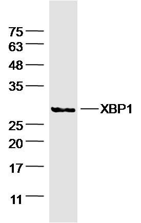
Sample: Liver(Mouse) Lysate at 40 ug
Primary: Anti- XBP1 (SL1668R) at 1/300 dilution
Secondary: IRDye800CW Goat Anti-Rabbit IgG at 1/20000 dilution
Predicted band size: 29kD
Observed band size: 29 kD
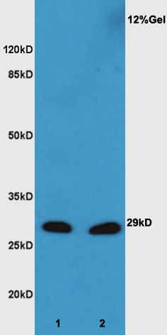
Sample:
Lane1: Embryo(Mouse) Lysate at 30 ug
Lane2: Liver (Mouse) Lysate at 30 ug
Primary: Anti-XBP-1(SL1668R) at 1:300 dilution;
Secondary: HRP conjugated Goat Anti-Rabbit IgG(SL0295G-HRP) at 1: 5000 dilution;
Predicted band size : 29kD
Observed band size : 29kD
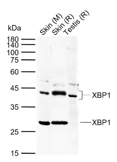
Sample:
Lane 1: Mouse Skin tissue lysates
Lane 2: Rat Skin tissue lysates
Lane 3: Rat Testis tissue lysates
Primary: Anti-XBP1 (SL1668R) at 1/500 dilution
Secondary: IRDye800CW Goat Anti-Rabbit IgG at 1/20000 dilution
Predicted band size: 29/40 kDa
Observed band size: 29/42 kDa
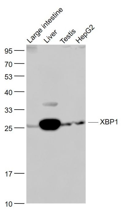
Sample:
Large intestine (Mouse) Lysate at 40 ug
Liver (Mouse) Lysate at 40 ug
Testis (Mouse) Lysate at 40 ug
HepG2(Human) Cell Lysate at 30 ug
Primary: Anti- XBP1 (SL1668R) at 1/1000 dilution
Secondary: IRDye800CW Goat Anti-Rabbit IgG at 1/20000 dilution
Predicted band size: 29/40 kD
Observed band size: 29 kD
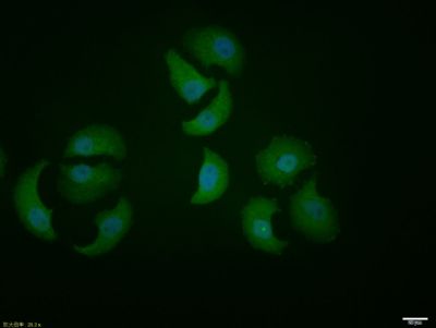
HepG2 cell; 4% Paraformaldehyde-fixed; Triton X-100 at room temperature for 20 min; Blocking buffer (normal goat serum, SLC0005) at 37°C for 20 min; Antibody incubation with (XBP1) polyclonal Antibody, Unconjugated (SL1668R) 1:100, 90 minutes at 37°C; followed by a conjugated Goat Anti-Rabbit IgG antibody at 37°C for 90 minutes, DAPI (blue, C02-04002) was used to stain the cell nuclei.
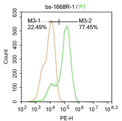
U-937 cells were fixed with 4% PFA for 10min at room temperature,permeabilized with 90% ice-cold methanol for 20 min at room temperature,and incubated in 5% BSA blocking buffer for 30 min at room temperature. Cells were then stained with XBP1 Antibody(SL1668R) at 1:500 dilution in blocking buffer and incubated for 30 min at room temperature, washed twice with 2%BSA in PBS, followed by secondary antibody incubation for 40 min at room temperature. Acquisitions of 20,000 events were performed.Cells stained with primary antibody (green), and isotype control (orange).
Product Feedback Wall
Message :
Your Email :
Copyright © 2007-2018 Sunlong Medical All Rights Reserved.