| Picture |
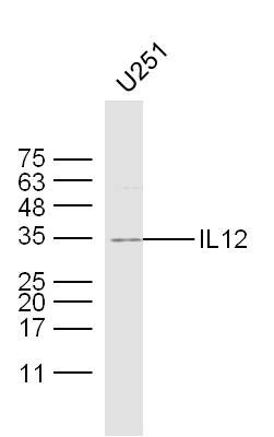
Sample: U251 Cell Lysate at 30 ug
Primary: Anti- IL12 (SL1789R) at 1/300 dilution
Secondary: IRDye800CW Goat Anti-Rabbit IgG at 1/20000 dilution
Predicted band size: 23 kD
Observed band size: 34 kD
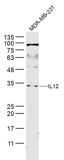
Sample:
MDA-MB-231(Human) Cell Lysate at 40 ug
Primary: Anti-IL12 (SL1789R) at 1/300 dilution
Secondary: IRDye800CW Goat Anti-Rabbit IgG at 1/20000 dilution
Predicted band size: 23 kD
Observed band size: 34 kD
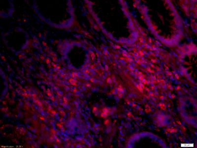
Tissue/cell: human colon carcinoma;4% Paraformaldehyde-fixed and paraffin-embedded;
Antigen retrieval: citrate buffer ( 0.01M, pH 6.0 ), Boiling bathing for 15min; Blocking buffer (normal goat serum,SLC0005) at 37∩ for 20 min;
Incubation: Anti-IL12 Polyclonal Antibody, Unconjugated(SL1789R) 1:500, overnight at 4∑C; The secondary antibody was Goat Anti-Rabbit IgG, Cy3 conjugated(SL0295G-Cy3)used at 1:200 dilution for 40 minutes at 37∑C. DAPI(5ug/ml,blue,SLC0033) was used to stain the cell nuclei
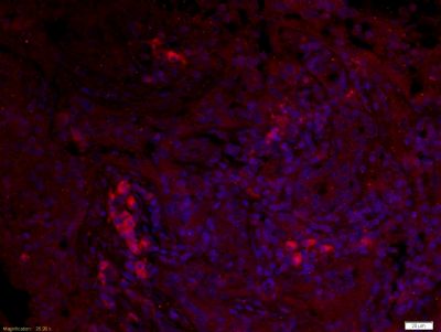
Tissue/cell: human lung carcinoma;4% Paraformaldehyde-fixed and paraffin-embedded;
Antigen retrieval: citrate buffer ( 0.01M, pH 6.0 ), Boiling bathing for 15min; Blocking buffer (normal goat serum,SLC0005) at 37∩ for 20 min;
Incubation: Anti-IL12 Polyclonal Antibody, Unconjugated(SL1789R) 1:500, overnight at 4∑C; The secondary antibody was Goat Anti-Rabbit IgG, Cy3 conjugated(SL0295G-Cy3)used at 1:200 dilution for 40 minutes at 37∑C. DAPI(5ug/ml,blue,SLC0033) was used to stain the cell nuclei
|