[IF=2.666] X Zhou et al. Modulating NMDA receptors to treat MK-801-induced schizophrenic cognition deficit: effects of clozapine combining with PQQ treatment and possible mechanisms of action. BMC Psychiatry. 2020 Mar 6;20(1):106. WB ; rat.
[IF=3.565] Liu J. et al. Moxidectin induces Cytostatic Autophagic Cell Death of Glioma Cells through inhibiting the AKT/mTOR Signalling Pathway.. J Cancer. 2020 Aug;11(19):5802-5811 WB ; Rat.
[IF=4.803] Liqin An. et al. Bone Morphogenetic Protein 4 (BMP4) promotes hepatic glycogen accumulation and reduces glucose level in hepatocytes through mTORC2 signaling pathway. Genes Dis. 2020 Nov;: WB,IHC ; Mouse.
[IF=10.679] Pan J et al. lncRNA JPX/miR-33a-5p/Twist1 axis regulates tumorigenesis and metastasis of lung cancer by activating Wnt/β-catenin signaling. Mol Cancer. 2020 Jan 15;19(1):9. WB ; Human.
[IF=2.84] Geng, Xiaofang, et al. "Differential proteome analysis of the cell differentiation regulated by BCC, CRH, CXCR4, GnRH, GPCR, IL1 signaling pathways in Chinese fire-bellied newt limb regeneration." Differentiation (2014). WB ;
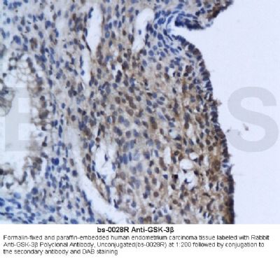
[IF=2.04] Gao, Hong, et al. "Comparative study of Hsp27, GSK3β, Wnt1 and PRDX3 in Hirschsprungs disease." International Journal of Experimental Pathology (2014). WB ; Human.
[IF=2.97] Li, Lingrui, et al. "Nrf2/ARE pathway activation, HO-1 and NQO1 induction by polychlorinated biphenyl quinone is associated with reactive oxygen species and PI3K/AKT signaling." Chemico-Biological Interactions (2013). WB ; Human.
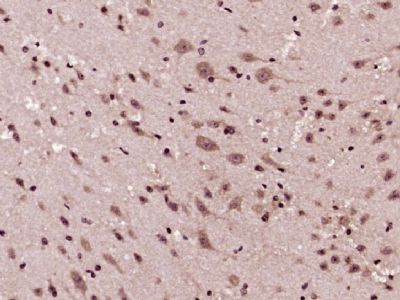
[IF=5.108] Yuan C et al. OAB-14, a bexarotene derivative, improves Alzheimer's disease-related pathologies and cognitive impairments by increasing β-amyloid clearance in APP/PS1 mice.(2019) Biochim Biophys Acta Mol Basis Dis.Jan;1865(1):161-36. WB ; Mouse.
[IF=2.884] Sun L et al. MiR-26a promotes fracture healing of nonunion rats possibly by targeting SOSTDC1 and further activating Wnt/β-catenin signaling pathway. Mol Cell Biochem. 2019 Jul 16. WB ; Rat.
[IF=3.04] Ma W et al. A vanillin derivative suppresses the growth of HT29 cells through the Wnt/β-catenin signaling pathway. Eur J Pharmacol. 2019 Apr 15;849:43-49. WB ; Human.
[IF=2.766] Freese et al. A novel blood-brain barrier co-culture system for drug targeting of Alzheimer's disease: establishment by using acitretin as a model drug. (2014) PLoS.One. 9:e91003 WB ; Human.
[IF=2.766] Zhang et al. Over-Expressed Twist Associates with Markers of Epithelial Mesenchymal Transition and Predicts Poor Prognosis in Breast Cancers via ERK and Akt Activation. (2015) PLoS.One. 10:e0135851 WB ; Human.
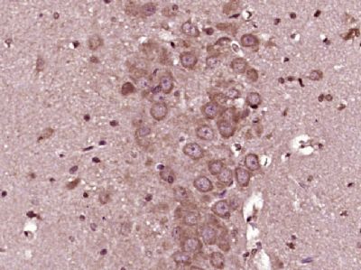
[IF=3.33] Zhao, Hai-hua, et al. "Involvement of GSK3 and PP2A in ginsenoside Rb1's attenuation of aluminum-induced tau hyperphosphorylation." Behavioural Brain Research (2012). WB ; Mouse.
[IF=3.7] Lin, Lai-xiang, et al. "Feasibility of β-Sheet Breaker Peptide-H102 Treatment for Alzheimers Disease Based on β-Amyloid Hypothesis." PLoS one 9.11 (2014): e112052. IHSLCP ; Mouse.