Integrins are important extracellular matrix (ECM) receptor proteins located on cell surfaces. They are hetrodimers composed of an alpha and a beta transmembrane glycoprotein subunit. Around twenty two different integrins (different alpha/ beta subunit combinations) are found in nature. Integrins are generally present in high concentrations at the cell surface, but, unlike most other cell surface receptors, they bind ligands with very low affinity. Due to their weak individual binding, integrins need to cluster and bind in groups in order to effectively bind the ECM. Integrins bind many different ligands including laminin. Each integrin is made up of a large N terminal extracellular domain that binds the ECM ligand and a small C terminal cytoplasmic domain that mediates interaction with the actin cytoskeleton and signaling function. Alpha 1 integrin along with alpha 2, alpha L and alpha M has a unique inserted domain. Integrin alpha 1 is a receptor for laminin and collagen. The alpha1 subunit is also known as CD49a. CD49a associates with CD29 (beta 1 integrin), to form an alpha1 beta1 heterodimer, identified as the rat homologue to VLA1, which is involved in cellular adhesion to laminin and collagen.
Function:
Integrin alpha-1/beta-1 is a receptor for laminin and collagen. It recognizes the proline-hydroxylated sequence G-F-P-G-E-R in collagen.
Subunit:
Heterodimer of an alpha and a beta subunit. Alpha-1 associates with beta-1. Interacts with RAB21.
Subcellular Location:
Membrane; Single-pass type I membrane protein.
Similarity:
Belongs to the integrin alpha chain family.
Contains 7 FG-GAP repeats.
Contains 1 VWFA domain.
SWISS:
P56199
Gene ID:
3672
Database links:
Entrez Gene: 3672 Human
Entrez Gene: 109700 Mouse
Entrez Gene: 25118 Rat
Omim: 192968 Human
SwissProt: P56199 Human
SwissProt: Q3V3R4 Mouse
SwissProt: P18614 Rat
Unigene: 644352 Human
Unigene: 482186 Mouse
Unigene: 91044 Rat
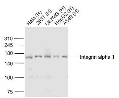
| Picture |
Sample:
Lane 1: Hela (Human) Cell Lysate at 30 ug
Lane 2: 293T (Human) Cell Lysate at 30 ug
Lane 3: U87MG (Human) Cell Lysate at 30 ug
Lane 4: HepG2 (Human) Cell Lysate at 30 ug
Lane 5: A549 (Human) Cell Lysate at 30 ug
Primary: Anti-Integrin alpha 1 (SL2095R) at 1/1000 dilution
Secondary: IRDye800CW Goat Anti-Rabbit IgG at 1/20000 dilution
Predicted band size: 150 kD
Observed band size: 160 kD
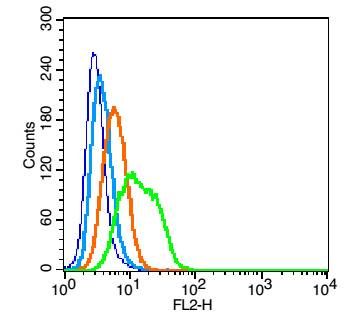
Blank control: U937(blue).
Primary Antibody: Rabbit Anti- Integrin alpha 1 antibody(SL2095R), Dilution: 1μg in 100 μL 1X PBS containing 0.5% BSA;
Isotype Control Antibody: Rabbit IgG(orange) ,used under the same conditions );
Secondary Antibody: Goat anti-rabbit IgG-PE(white blue), Dilution: 1:200 in 1 X PBS containing 0.5% BSA.
Protocol
The cells were fixed with 2% paraformaldehyde (10 min). Primary antibody (SL2095R, 1μg /1x10^6 cells) were incubated for 30 min on the ice, followed by 1 X PBS containing 0.5% BSA + 1 0% goat serum (15 min) to block non-specific protein-protein interactions. Then the Goat Anti-rabbit IgG/PE antibody was added into the blocking buffer mentioned above to react with the primary antibody at 1/200 dilution for 30 min on ice. Acquisition of 20,000 events was performed.
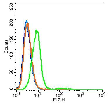
Blank control: RSC96 cells(blue).
Primary Antibody: Rabbit Anti-Integrin alpha 1 antibody(SL2095R), Dilution: 5μg in 100 μL 1X PBS containing 0.5% BSA;
Isotype Control Antibody: Rabbit IgG (orange) ,used under the same conditions.
Secondary Antibody: Goat anti-rabbit IgG-PE(white blue), Dilution: 1:200 in 1 X PBS containing 0.5% BSA.
|
|
|