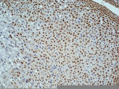
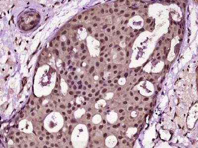
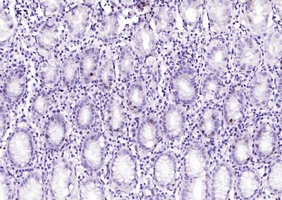
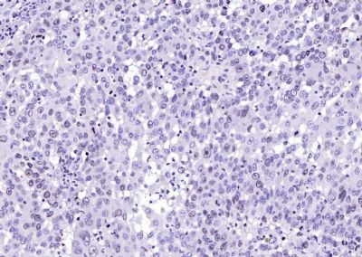
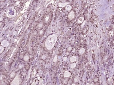
[IF=2.303] Suihui Li. et al. Hsa_circ_0048674 facilitates hepatocellular carcinoma progression and natural killer cell exhaustion depending on the regulation of miR-223-3p/PDL1. Histol Histopathol. 2022 Feb 21;1888 IHC ; Mouse.
[IF=5.858] Rong Fu. et al. Avenanthramide C induces cellular senescence in colorectal cancer cells via suppressing β-catenin-mediated the transcription of miR-183/96/182 cluster. Biochem Pharmacol. 2022 May;199:115021 IHC ; Human.
[IF=5.168] Dong et al. YY1 promotes HDAC1 expression and decreases sensitivity of hepatocellular carcinoma cells to HDAC inhibitor. (2017) Oncotarget. 8:40583-40593 IHSLCP ; Human.
[IF=1.438] Jiang JY et al. Effects of estradiol and progesterone on secretion of epidermal growth factor and insulin-like growth factor-1 in cultured yak endometrial epithelial cells. Tissue Cell. 2018 Jun;52:28-34. IF&WB ; Yak.
[IF=5.959] Hou Y et al. SKA3 Promotes tumor growth by regulating CDK2/P53 phosphorylation in hepatocellular carcinoma. Cell Death Dis. 2019 Dec 5;10(12):929. IHC ; Human.
[IF=2.906] Jinfeng Zhu et al. Expression and prognostic significance of pyruvate dehydrogenase kinase 1 in bladder urothelial carcinoma. Virchows Arch. 2020 Nov;477(5):637-649. IHC ; Human.
[IF=5.026] Di Xia et al. Nrf2 promotes esophageal squamous cell carcinoma (ESCC) resistance to radiotherapy through the CaMKIIα-associated activation of autophagy. Cell Biosci
. 2020 Jul 30;10:90. IHC ; Human.
[IF=14.593] Guo-Bin Ding. et al. Molecularly engineered tumor acidity-responsive plant toxin gelonin for safe and efficient cancer therapy. Bioact Mater. 2022 Feb;: IHC ; Mouse.
[IF=5.279] Peng Yang. et al. Cucurbitacin E Triggers Cellular Senescence in Colon Cancer Cells via Regulating the miR-371b-5p/TFAP4 Signaling Pathway. J Agr Food Chem. 2022;XXXX(XXX):XXX-XXX IHC ; Mouse.
[IF=6.4] Chu, Min. et al. Knockdown of lncRNA BDNF-AS inhibited the progression of multiple myeloma by targeting the miR-125a/b-5p-BCL2 axis. Immun Ageing. 2022 Dec;19(1):1-17 IHC ; Mouse.
[IF=10.435] Wan, Guoyun. et al. Gene augmented nuclear-targeting sonodynamic therapy via Nrf2 pathway-based redox balance adjustment boosts peptide-based anti-PD-L1 therapy on colorectal cancer. J Nanobiotechnol. 2021 Dec;19(1):1-26 IF ; Mouse.
[IF=5.115] Liang Kong. et al. Tumor Microenvironmental Responsive Liposomes Simultaneously Encapsulating Biological and Chemotherapeutic Drugs for Enhancing Antitumor Efficacy of NSCLC. Int J Nanomed. 2020; 15: 6451–6468 IHC ; Human.
[IF=3.337] Zhaohui Jia. et al. LncRNA MCM3AP-AS1 Promotes Cell Proliferation and Invasion Through Regulating miR-543-3p/SLC39A10/PTEN Axis in Prostate Cancer. Oncotargets Ther. 2020; 13: 9365–9376 IHC ; Human.
[IF=3.082] Jingyi You. et al. Human Umbilical Cord Mesenchymal Stem Cell-Derived Small Extracellular Vesicles Alleviate Lung Injury in Rat Model of Bronchopulmonary Dysplasia by Affecting Cell Survival and Angiogenesis. Stem Cells Dev. 2020 Nov;29(23):1520-1532 IF ; Rat.
[IF=6.304] Feng Y et al. Long noncoding RNA HOXA-AS2 functions as an oncogene by binding to EZH2 and suppressing LATS2 in acute myeloid leukemia (AML)Cell Death Dis.2020 Dec 2;11(12):1025. IHC ; Mouse.
[IF=5.115] Xia Qinet al. Zinc Oxide Nanoparticles Induce Ferroptotic Neuronal Cell Death in vitro and in vivo. Int J Nanomedicine
. 2020 Jul 27;15:5299-5315. IF ; mouse.
[IF=3.647] Lin D et al. Aldehyde dehydrogenase 2 regulates autophagy via the Akt-mTOR pathway to mitigate renal ischemia-reperfusion injury in hypothermic machine perfusion. Life Sci
. 2020 Jul 15;253:117705. IHC,WB ; mouse.
[IF=8.355] Xie S et al. Bacteria-propelled microrockets to promote the tumor accumulation and intracellular drug uptake. Chemical Engineering Journal,2019, 123786. IHC ; Mouse.
[IF=2.705] Zhang Q et al. The circular RNA hsa_circ_0007623 acts as a sponge of microRNA-297 and promotes cardiac repair. Biochem Biophys Res Commun. 2020 Mar 19;523(4):993-1000. IHSLCP ; Mouse.
[IF=3.743] Liu F et al. Clinical and biological significances of heat shock protein 90 (Hsp90) in human nasopharyngeal carcinoma cells and anti-cancer effects of Hsp90 inhibitor. Biomed Pharmacother. 2019 Oct 18;120:109533. ICF ; Human.
[IF=3.598] Li R et al. Pharmacological biotargets and the molecular mechanisms of oxyresveratrol treating colorectal cancer: Network and experimental analyses. Biofactors. 2019 Oct 24. IHF&ICF ; Human.
[IF=3.06] Li T et al. Withanolides, extracted from Datura metel L. inhibit keratinocyte proliferation and imiquimod- induced psoriasis-like dermatitis via the STAT3/P38/ERK1/2 pathway. Molecules. 2019 Jul 17;24(14). pii: E2596. WB ; Mouse.
[IF=2.663] Tang Y et al. LncRNA differentiation antagonizing non-protein coding RNA promotes proliferation and invasion through regulating miR-135a/NLRP37 axis in pancreatic cancer. Invest New Drugs. 2019 Jul 3. IHC ; Mouse.
[IF=8.355] Chen Z et al. Enzyme-powered Janus nanomotors launched from intratumoral depots to address drug delivery barriers. Chemical Engineering Journal,2019 375, 122109. IHC ; Mouse.
[IF=5.047] Chen Z et al. Synergistic antitumor efficacy of hybrid micelles with mitochondrial targeting and stimuli-responsive drug release. Journal of Materials Chemistry B.2019. IHSLCP ; Human.
[IF=6.383] Xie S et al. Bacterial microbots for acid-labile release of hybrid micelles to promote the synergistic antitumor efficacy.Acta Biomater. 2018 Sep 15;78:198-210. IHSLCP ; Mouse.
[IF=3.923] Sha Y et al. MGF E peptide improves anterior cruciate ligament repair by inhibiting hypoxia‐induced cell apoptosis and accelerating angiogenesis.(2018) J Cell Physiol. 2018 Oct 14. IF ; Rabbit.
[IF=1.39] Wu et al. A retrospective, single-center cohort study on 65 patients with primary retroperitoneal liposarcoma. (2018) Oncol.Lett. 15:1799-1810 IHSLCP ; Human.
[IF=2.766] Li et al. Visfatin Destabilizes Atherosclerotic Plaques in Apolipoprotein E-Deficient Mice. (2016) PLoS.One. 11:e0148273 WB ; Rat.
[IF=4.12] Zheng et al. The Modification of Tet1 in Male Germline Stem Cells and Interact with PCNA, HDAC1 to promote their Self-renewal and Proliferation. (2016) Sci.Rep. 6:37414 IF ; Goat.
[IF=7.504] He, Nan, et al. "Tumor pH-responsive Release of Drug-conjugated Micelles from Fiber Fragments for Intratumoral Chemotherapy." ACS Applied Materials & Interfaces (2017). other ;
[IF=3.54] Thompson, Christopher, et al. "Loss of caveolin-1 alters extracellular matrix protein expression and ductal architecture in murine mammary glands." PLOS ONE 12.2 (2017): e0172067. IHSLCP ; Mouse.
[IF=0.66] Aslaner, Arif, et al. "Medical ozone treatment ameliorates the acute distal colitis in rat." Acta Cirurgica Brasileira 31.4 (2016): 256-263. IHSLCP ; Rat.
[IF=5.04] Drel, Viktor R., et al. "Centrality of bone marrow in the severity of gadolinium-based contrast-induced systemic fibrosis." The FASEB Journal(2016): fj-20300188R. IHSLCP ; Rat.
[IF=1.27] Ma, Wei‑Yuan, Kun Jia, and Yan Zhang. "IL-17 promotes keratinocyte proliferation via the downregulation of C/EBPα." Experimental and Therapeutic Medicine. IHSLCP ; Human.
[IF=2.43] Zhang, Jing-Yao, et al. "Hydrogen-rich water protects against acetaminophen-induced hepatotoxicity in mice." World Journal of Gastroenterology 21.14 (2015): 4195-4209. IHSLCP ; Mouse.
[IF=8.31] Pan, Dayi, et al. "PEGylated dendritic diaminocyclohexyl-platinum (II) conjugates as pH-responsive drug delivery vehicles with enhanced tumor accumulation and antitumor efficacy." Biomaterials (2014). IHSLCP ; Mouse.
[IF=4.75] Zhao, Yuechao, et al. "Estrogen-Induced CCN1 is Critical for Establishment of Endometriosis-like Lesions in Mice." Molecular Endocrinology (2014). IHSLCP ; Mouse.
[IF=4.2] Yuan, Qing, et al. "Docetaxel-loaded solid lipid nanoparticles suppress breast cancer cells growth with reduced myelosuppression toxicity." International Journal of Nanomedicine 9 (2014): 4829. WB ; Mouse.
[IF=2.07] Ma, Ying-Yu, et al. "Involvement of c-KIT mutation in the development of gastrointestinal stromal tumors through proliferation promotion and apoptosis inhibition." OncoTargets and Therapy 7 (2014): 637-643. IHSLCP ; Human.
[IF=6.03] Li, Xin, Xiumei Zhu, and Liyan Qiu. "Constructing aptamer anchored nanovesicles for enhanced tumor penetration and cellular uptake of water soluble chemotherapeutics." Acta Biomaterialia (2016). IHSLCP ; Mouse.
[IF=3.269] Gongjun Xu. et al. Expression and significance of mammalian target of rapamycin in cutaneous squamous cell carcinoma and precancerous lesions. Bioengineered. 2021;12(2):9930-9938 IHC ; Human.
[IF=11.161] Han, Lili. et al. The RNA-binding protein GRSF1 promotes hepatocarcinogenesis via competitively binding to YY1 mRNA with miR-30e-5p. J Exp Clin Canc Res. 2022 Dec;41(1):1-18 IHC ; Mouse.
[IF=7.658] Hua-Li Wang. et al. Potential Synthetic Lethality for Breast Cancer: A Selective Sirtuin 2 Inhibitor Combined with a Multiple Kinase Inhibitor Sorafenib. Pharmacol Res. 2021 Dec;:106050 IF,IHC ; Mouse.
[IF=5.241] Zhou, Guanwen. et al. FTO promotes tumour proliferation in bladder cancer via the FTO/miR-576/CDK6 axis in an m6A-dependent manner. Cell Death Discov. 2021 Nov;7(1):1-11 IHC ; Human.
[IF=13.273] Long Zhao. et al. Juglone-loaded metal-organic frameworks for H2O2 self-modulating enhancing chemodynamic therapy against prostate cancer. Chem Eng J. 2022 Feb;430:133057 IHC ; Mouse.
[IF=5.414] Wenhuan Guo. et al. Stem cells from human exfoliated deciduous teeth affect mitochondria and reverse cognitive decline in a senescence-accelerated mouse prone 8 model. Cytotherapy. 2021 Sep;: IF,IHC ; mouse.
[IF=4.534] Chao Yang. et al. Circ_0006988 promotes the proliferation, metastasis and angiogenesis of non-small cell lung cancer cells by modulating miR-491-5p/MAP3K3 axis. 2021 Jun 30 IHC ; Mouse.
[IF=3.082] Jiang Liu. et al. Type 2 Alveolar Epithelial Cells Differentiated from Human Umbilical Cord Mesenchymal Stem Cells Alleviate Mouse Pulmonary Fibrosis through β-Catenin-Regulated Cell Apoptosis. 2021 Apr 24 IHC ; Mouse.
[IF=13.116] Dongyang Zhao. et al. Apoptotic body–mediated intercellular delivery for enhanced drug penetration and whole tumor destruction. Sci Adv. 2021 Apr;7(16):eabg0176 IHC ; Mouse.
[IF=14.588] Donglin Xia. et al. Au–Hemoglobin Loaded Platelet Alleviating Tumor Hypoxia and Enhancing the Radiotherapy Effect with Low-Dose X-ray. Acs Nano. 2020;14(11):15654–15668 IHC ; Mouse.
[IF=4.175] Jia Guo. et al. Circ_0000140 restrains the proliferation, metastasis and glycolysis metabolism of oral squamous cell carcinoma through upregulating CDC73 via sponging miR-182-5p. Cancer Cell Int. 2020 Dec;20(1):1-12 WB ; Human.
[IF=3.15] Gong H et al. EZH2 inhibitors reverse resistance to gefitinib in primary EGFR wild-type lung cancer cellsBMC Cancer.2020 Dec 4;20(1):1189. IHC ; Human.
[IF=10.317] Qi Sun. et al. Phototherapy and anti-GITR antibody-based therapy synergistically reinvigorate immunogenic cell death and reject established cancers. Biomaterials. 2021 Feb;269:120648 IHC ; Mouse.
[IF=4.831] Chengjun Sunet al. Long noncoding RNA AC092171.4 promotes hepatocellular carcinoma progression by sponging microRNA-1271 and upregulating GRB2. Aging (Albany NY)
. 2020 Jul 21;12(14):14141-14156. IHC ; Human.
[IF=4.225] Jin Y et al. Oxymatrine Inhibits Renal Cell Carcinoma Progression by Suppressing β-Catenin Expression. Front Pharmacol
. 2020 Jun 5;11:808. IHSLCP ; Human.
