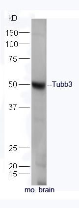
Sample: Brain(Mouse) lysate at 30ug;
Primary: Anti-Tubb3 (SL2670R) at 1:300 dilution;
Secondary: HRP conjugated Goat-Anti-rabbit IgG(SL0295G-HRP) at 1: 5000 dilution;
Predicted band size: 50 kD
Observed band size: 50 kD
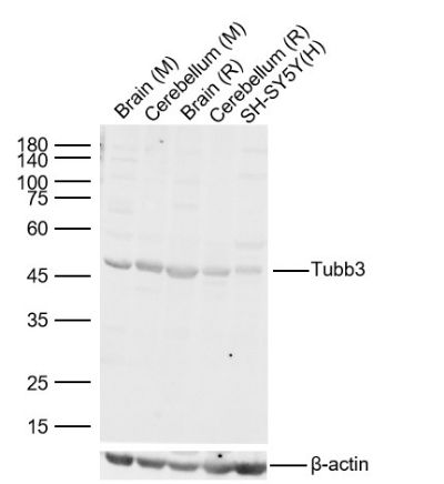
Sample:
Lane 1: Mouse Brain Lysates
Lane 2: Mouse Cerebellum Lysates
Lane 3: Rat Brain Lysates
Lane 4: Rat Cerebellum Lysates
Lane 5: Human SH-SY5Y cell Lysates
Primary: Anti-Tubb3 (SL2670R) at 1/2000 dilution
Secondary: IRDye800CW Goat Anti-Rabbit IgG at 1/20000 dilution
Predicted band size: 50kDa
Observed band size: 50 kDa
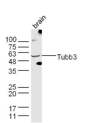
Sample: Brain(Mouse) lysate at 30ug;
Primary: Anti-Tubb3 (SL2670R) at 1:300 dilution;
Secondary: HRP conjugated Goat-Anti-rabbit IgG(SL0295G-HRP) at 1: 5000 dilution;
Predicted band size: 50 kD
Observed band size: 50 kD
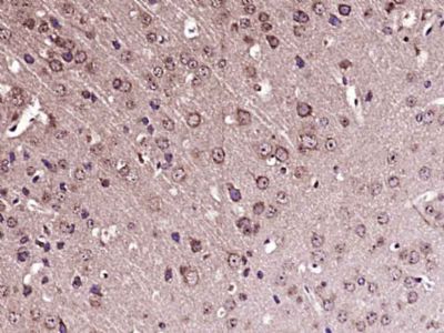
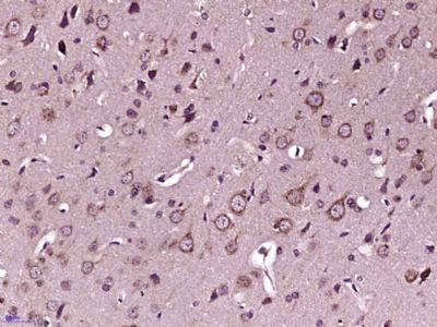
Paraformaldehyde-fixed, paraffin embedded (Mouse brain); Antigen retrieval by boiling in sodium citrate buffer (pH6.0) for 15min; Block endogenous peroxidase by 3% hydrogen peroxide for 20 minutes; Blocking buffer (normal goat serum) at 37°C for 30min; Antibody incubation with (Tubb3) Polyclonal Antibody, Unconjugated (SL2670R) at 1:400 overnight at 4°C, followed by operating according to SP Kit(Rabbit) (sp-0023) instructionsand DAB staining.
Paraformaldehyde-fixed, paraffin embedded (Rat brain); Antigen retrieval by boiling in sodium citrate buffer (pH6.0) for 15min; Block endogenous peroxidase by 3% hydrogen peroxide for 20 minutes; Blocking buffer (normal goat serum) at 37°C for 30min; Antibody incubation with (Tubb3) Polyclonal Antibody, Unconjugated (SL2670R) at 1:400 overnight at 4°C, followed by operating according to SP Kit(Rabbit) (sp-0023) instructionsand DAB staining.
|