[IF=4.171] Gu F et al. Lactobacillus casei improves depression-like behavior in chronic unpredictable mild stress-induced rats by BDNF-TrkB signal pathway and the intestinal microbiota. Food Funct
. 2020 Jul 1;11(7):6148-6157. WB ; Rat.
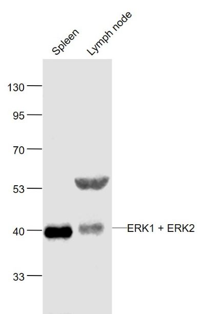
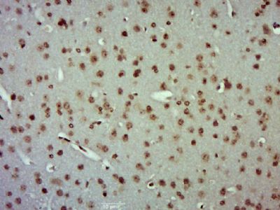
[IF=2.316] Song, Yue. et al. Seasonal expression of extracellular signal regulated kinases in the colon of wild ground squirrels (Spermophilus dauricus). Mol Biol Rep. 2022 Jan;:1-7 WB,IHC ; Squirrel.
[IF=3.352] Yusong Miao. et al. Methylsulfonylmethane ameliorates inflammation via NF-κB and ERK/JNK-MAPK signaling pathway in chicken trachea and HD11 cells during Mycoplasma gallisepticum infection. Poultry Sci. 2022 Jan;:101706 WB ; Chicken.
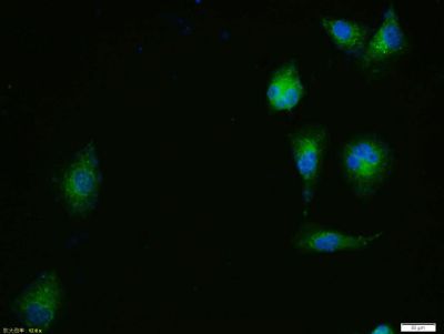
[IF=5.923] Junfeng Ke. et al. CTI-2 Inhibits Metastasis and Epithelial-Mesenchymal Transition of Breast Cancer Cells by Modulating MAPK Signaling Pathway. Int J Mol Sci. 2021 Jan;22(22):12229 WB,IF ; Human.
[IF=4.486] Jianye Yang. et al. Astragalus polysaccharide attenuates LPS-related inflammatory osteolysis by suppressing osteoclastogenesis by reducing the MAPK signalling pathway. 2021 Jun 02 WB ; Mouse.
[IF=4.105] Ai Jin. et al. TSLP-induced collagen type-I synthesis through STAT3 and PRMT1 is sensitive to calcitriol in human lung fibroblasts. Bba-Mol Cell Res. 2021 Jun;:119083 WB ; Human.
[IF=4.225] Qihe Tang. et al. Bergenin Monohydrate Attenuates Inflammatory Response via MAPK and NF-κB Pathways Against Klebsiella pneumonia Infection. Front Pharmacol. 2021; 12: 651664 WB ; Mouse.
[IF=3.647] Meixuan Chen. et al. Activation of the δ opioid receptor relieves cerebral ischemic injury in rats via EGFR transactivation. Life Sci. 2021 May;273:119292 WB ; Rat.
[IF=1.713] Jian Wang. et al. Lactobacillus salivarius ameliorated Mycoplasma gallisepticum-induced inflammatory injury and secondary Escherichia coli infection in chickens: Involvement of intestinal microbiota. Vet Immunol Immunop. 2021 Mar;233:110192 WB ; Chicken.
[IF=4.171] Gu F et al. Lactobacillus casei improves depression-like behavior in chronic unpredictable mild stress-induced rats by the BDNF-TrkB signal pathway and the intestinal microbiota. Food Funct. 2020 Jul 1;11(7):6148-6157. WB ; Rat.
[IF=3.743] Liu F et al. Clinical and biological significances of heat shock protein 90 (Hsp90) in human nasopharyngeal carcinoma cells and anti-cancer effects of Hsp90 inhibitor. Biomed Pharmacother. 2019 Oct 18;120:109533. ICF ; Human.
[IF=3.266] Ma Q et al. Vitamin B5 inhibit RANKL induced osteoclastogenesis and ovariectomy induced osteoporosis by scavenging ROS generation. Am J Transl Res. 2019 Aug 15;11(8):5008-5018. eCollection 2019. WB ; Mouse.
[IF=2.394] Liu W et al. Synergistic effect of tolfenamic acid and glycyrrhizic acid on TPA-induced skin inflammation in mice. MedChemComm.2019. IHSLCP ; Mouse.
[IF=5.614] Zhu J et al. SPARC Promotes Self‐Renewal of Limbal Epithelial Stem Cells and Ocular Surface Restoration through JNK and p38‐MAPK Signaling Pathways. Stem Cells. 2019 Oct 23. WB ; Rabbit.
[IF=2.027] Li QH et al. Effect of heat stress on mitogen-activated protein kinases in the hypothalamic− pituitary− gonadal axis of developing Wenchang chicks. Poultry Science.2019. WB ; chick.
[IF=4.61] Yue H et al. Gestational exposure to PM2.5 impairs vascularization of the placenta. Sci Total Environ. 2019 May 15;665:153-161. WB ; Mouse.
[IF=3.249] Lin X et al. TFF3 Contributes to Epithelial-Mesenchymal Transition (EMT) in Papillary Thyroid Carcinoma Cells via the MAPK/ERK Signaling Pathway.(2018)J Cancer;Oct 31;9(23):4430-4439. ICC ; Human.
[IF=2.342] Liu W et al. Glycyrrhizic acid from licorice down-regulates inflammatory responses via blocking MAPK and PI3K/Akt-dependent NF-κB signalling pathways in TPA-induced skin inflammation. Medchemcomm. 2018 Jul 19;9(9):1502-1510. IHSLCP ; Mouse.
[IF=2.342] Liu W et al. Glycyrrhizic acid from licorice down-regulates inflammatory responses via blocking MAPK and PI3K/Akt-dependent NF-κB signalling pathways in TPA-induced skin inflammation. MedChemComm. 2018. IHSLCP ; Mouse.
[IF=1.374] Zheng et al. Ginsenoside Rb1 improves cardiac function and remodeling in heart failure. (2017) Exp.Ani. 66:217-228 WB ; Rat.
[IF=1.39] Li et al. Downregulation of BRAF-activated non-coding RNA suppresses the proliferation, migration and invasion, and induces apoptosis of hepatocellular carcinoma cells. (2017) Oncol.Lett. 14:4751-4757 WB ; Human.
[IF=4.2] Zhang, Fengwei, et al. "Seasonal Expression of Oxytocin and Oxytocin Receptor in the Scented Gland of Male Muskrat (Ondatra zibethicus)." Scientific Reports 7.1 (2017): 16627. WB, IHSLCP ; Others.
[IF=1.69] Liu, Xinwei, et al. "Inhibition of BTK protects lungs from trauma-hemorrhagic shock-induced injury in rats." Molecular Medicine Reports 16.1 (2017): 192-200. WB ; Rat.
[IF=4.42] Yu, Haijie, et al. "Gypenoside Protects Cardiomyocytes against Ischemia-Reperfusion Injury via the Inhibition of Mitogen-Activated Protein Kinase Mediated Nuclear Factor Kappa B Pathway In Vitro and In Vivo." Frontiers in Pharmacology 7 (2016). WB ; Rat.
[IF=3.53] Wang T, Zhao L, Guo Y, Zhang M, Pei H (2015) Picroside II Inhibits Neuronal Apoptosis and Improves the Morphology and Structure of Brain Tissue following Cerebral Ischemic Injury in Rats. PLoS ONE 10(4): e014899. IHSLCP ; Rat.
[IF=2.41] Xu, Shan, et al. "Acyldepsipeptides inhibit the growth of renal cancer cells through G1 phase cell cycle arrest." Biochemical and Biophysical Research Communications (2013). WB ; Human.
[IF=2.38] Cong, Lin, and Wenting Chen. "Neuroprotective Effect of Ginsenoside Rd on Spinal Cord Injury Rats." Basic & Clinical Pharmacology & Toxicology(2016). WB ; Rat.
[IF=3.738] Lili Liu. et al. Rutin Ameliorates Cadmium-Induced Necroptosis in the Chicken Liver via Inhibiting Oxidative Stress and MAPK/NF-κB Pathway. 2021 Jun 06 WB ; Chicken.
[IF=1.713] Jian Wang. et al. A respiratory commensal bacterium acts as a risk factor for Mycoplasma gallisepticum infection in chickens. Vet Immunol Immunop. 2020 Dec;230:110127 WB ; Chicken.
[IF=4.556] Dongyi Xu. et al. The Critical Role of NLRP6 Inflammasome in Streptococcus pneumoniae Infection In Vitro and In Vivo. Int J Mol Sci. 2021 Jan;22(8):3876 WB ; Mouse.
[IF=3.641] Wenbo Ge. et al. 17β-estradiol protects sheep oviduct epithelial cells against lipopolysaccharide-induced inflammation in vitro. Mol Immunol. 2020 Nov;127:21 WB ; Sheep.
[IF=2.445] Yuan Z et al. Seasonal expressions of oxytocin and oxytocin receptor in the epididymides in the wild ground squirrels (Citellus Dauricus Brandt). Gen Comp Endocrinol. 2020 Apr 1;289:113391. WB&IHSLCP ; Wild ground squirrel.
[IF=1.719] Zhou J et al. Paeonol antagonizes oncogenesis of osteosarcoma by inhibiting the function of TLR4/MAPK/NF-κB pathway. Acta Histochem. 2019 Oct 3:151455. WB ; Mouse&Human.
[IF=4.658] Zhang J et al. Down‐regulation of Suv39h1 attenuates neointima formation after carotid artery injury in diabetic rats. J Cell Mol Med. 2019 Nov 17. WB ; Rat.
[IF=5.391] Shin WS et al. Catalytically inactive receptor tyrosine kinase PTK7 activates FGFR1 independent of FGF. FASEB J. 2019 Sep 18:fj201900932R. WB ; Human.
[IF=4.183] Imoto K et al. Periostin Mediates Right Ventricular Failure through Induction of Inducible Nitric Oxide SynthaseExpression in Right Ventricular Fibroblasts from Monocrotaline-Induced Pulmonary Arterial Hypertensive Rats. Int J Mol Sci. 2018 Dec 24;20(1). pii: E62. IHSLCP ; Rat.
[IF=0] Cong and Chen Neuroprotective Effect of Ginsenoside Rd on Spinal Cord Injury Rats. (2016) Basic.Clin.Pharmacol.Toxicol. WB ; Rat.
[IF=0.764] Chen et al. Downregulation of MACC1 expression enhances cisplatin sensitivity in SKOSLV3/DDP cells. (2015) Genet.Mol.Re. 14:17134-44 WB ; Human.
[IF=1.39] Li, Jing, et al. "Downregulation of BRAF‑activated non‑coding RNA suppresses the proliferation, migration and invasion, and induces apoptosis of hepatocellular carcinoma cells." Oncology Letters 14.4 (2017): 4751-4757. WB ; Human.
[IF=2.55] Zhao, Haiyan, et al. "Inhibition of endocan attenuates monocrotaline-induced connective tissue disease related pulmonary arterial hypertension." International Immunopharmacology 42 (2017): 115-121. WB ; Rat.
[IF=3.04] Chen, Wei, et al. "Mycobacterium tuberculosis PE25/PPE41 protein complex induces activation and maturation of dendritic cells and drives Th2-biased immune responses." Medical Microbiology and Immunology (2015): 1-13. WB ; Mouse.
[IF=2.47] Zhao, Hongyu, et al. "Betulin attenuates lung and liver injuries in sepsis."International Immunopharmacology 30 (2016): 50-56. WB ; Rat.
[IF=2.4] Wang, Tingting, et al. "Picroside II has a neuroprotective effect by inhibiting ERK1/2 activation after cerebral ischemic injury in rats." Clinical and Experimental Pharmacology and Physiology (2015). IHSLCP ; Rat.
[IF=2.75] Dang, Hui-Min, et al. "“Qufeng Tongluo” acupuncture Prevents the progression of glomerulonephritis by Decreasing renal sympathetic nerve activity." Journal of Ethnopharmacology (2014). WB ; Rabbit.