Home > Product > Antibody > Rabbit Anti-phospho-MCSF Receptor (Tyr923) antibody
MCSF Receptor; C FMS; CD 115; CD115; CD115 antigen; CFMS; Colony stimulating factor 1 receptor; Colony stimulating factor I receptor; CSF 1 R; CSF 1R; CSF-1 receptor; CSF-1-R; CSF1 R; CSF1R; CSF1R_HUMAN; CSFR; FIM 2; FIM2; FMS; FMS proto oncogene; FMS pro
Cat:
SL3079R
Species Reactivity:
Human,Mouse,
Immunogen:
KLH conjugated Synthesised phosphopeptide derived from human CSF1R around the phosphorylation site of Tyr923:RD(p-Y)TN<Cytoplasmic>
Format:
Liquid
Storage instructions:
Shipped at 4℃. Store at -20 °C for one year. Avoid repeated freeze/thaw cycles.
Concentration:
1mg/ml
Clonality:
Polyclonal
Isotype:
IgG
Applications:
ELISA=1:5000-10000not yet tested in other applications.optimal dilutions/concentrations should be determined by the end user.
Host:
Rabbit
Product Overview:
Blank control: Raw264.7. Primary Antibody (green line): Rabbit Anti-phospho-MCSF Receptor (Tyr923) antibody (SL3079R) Dilution: 1μg /10^6 cells; Isotype Control Antibody (orange line): Rabbit IgG . Secondary Antibody : Goat anti-rabbit IgG-AF647Dilution: 1μg /test. ProtocolThe cells were incubated in 5%BSA to block non-specific protein-protein interactions for 30 min at room temperature .Cells stained with Primary Antibody for 30 min at room temperature. The secondary antibody used for 40 min at room temperature. Acquisition of 20,000 events was performed.Blank control (blue line): MCF7 (blue). Primary Antibody (green line): Rabbit Anti-phospho-MCSF Receptor(Tyr923) antibody (SL3079R) Dilution: 1μg /10^6 cells; Isotype Control Antibody (orange line): Rabbit IgG . Secondary Antibody (white blue line): F(ab’)2 fragment goat anti-rabbit IgG-FITCDilution: 1μg /test. ProtocolThe cells were fixed with 2% paraformaldehyde for 10 min at room temperature.Cells stained with Primary Antibody for 30 min at room temperature. The cells were then incubated in 1 X PBS/2%BSA/10% goat serum to block non-specific protein-protein interactions followed by the antibody for 15 min at room temperature. The secondary antibody used for 40 min at room temperature. Acquisition of 20,000 events was performed.
More
Unit:
Price: $
Product PDFs
Datasheet:


This protein tyrosine kinase transmembrane receptor is the receptor for colony stimulating factor 1, a cytokine which controls the production, differentiation, and function of macrophages. This receptor mediates most if not all of the biological effects of this cytokine. Ligand binding activates the receptor kinase through a process of oligomerization and transphosphorylation. The encoded protein is a member of the CSF1/PDGF receptor family of tyrosine protein kinases and contains 5 immunoglobulin like C2 type domains. CD115 is expressed by cells of the monocytic lineage and by progenitor cells. Mutations in this gene have been associated with a predisposition to myeloid malignancy.

Function:
Tyrosine-protein kinase that acts as cell-surface receptor for CSF1 and IL34 and plays an essential role in the regulation of survival, proliferation and differentiation of hematopoietic precursor cells, especially mononuclear phagocytes, such as macrophages and monocytes. Promotes the release of proinflammatory chemokines in response to IL34 and CSF1, and thereby plays an important role in innate immunity and in inflammatory processes. Plays an important role in the regulation of osteoclast proliferation and differentiation, the regulation of bone resorption, and is required for normal bone and tooth development. Required for normal male and female fertility, and for normal development of milk ducts and acinar structures in the mammary gland during pregnancy. Promotes reorganization of the actin cytoskeleton, regulates formation of membrane ruffles, cell adhesion and cell migration, and promotes cancer cell invasion. Activates several signaling pathways in response to ligand binding. Phosphorylates PIK3R1, PLCG2, GRB2, SLA2 and CBL. Activation of PLCG2 leads to the production of the cellular signaling molecules diacylglycerol and inositol-1,4,5-trisphosphate, that then lead to the activation of protein kinase C family members, especially PRKCD. Phosphorylation of PIK3R1, the regulatory subunit of phosphatidylinositol 3-kinase, leads to activation of the AKT1 signaling pathway. Activated CSF1R also mediates activation of the MAP kinases MAPK1/ERK2 and/or MAPK3/ERK1, and of the SRC family kinases SRC, FYN and YES1. Activated CSF1R transmits signals both via proteins that directly interact with phosphorylated tyrosine residues in its intracellular domain, or via adapter proteins, such as GRB2. Promotes activation of STAT family members STAT3, STAT5A and/or STAT5B. Promotes tyrosine phosphorylation of SHC1 and INPP5D/SHIP-1. Receptor signaling is down-regulated by protein phosphatases, such as INPP5D/SHIP-1, that dephosphorylate the receptor and its downstream effectors, and by rapid internalization of the activated receptor.

Subunit:
Interacts with INPPL1/SHIP2 and THOC5. Monomer. Homodimer. Interacts with CSF1 and IL34. Interaction with dimeric CSF1 or IL34 leads to receptor homodimerization. Interacts (tyrosine phosphorylated) with PLCG2 (via SH2 domain). Interacts (tyrosine phosphorylated) with PIK3R1 (via SH2 domain). Interacts (tyrosine phosphorylated) with FYN, YES1 and SRC (via SH2 domain). Interacts (tyrosine phosphorylated) with CBL, GRB2 and SLA2. .

Subcellular Location:
Cell membrane; Single-pass type I membrane protein.

Tissue Specificity:
Expressed in bone marrow and in differentiated blood mononuclear cells.

Post-translational modifications:
Autophosphorylated in response to CSF1 or IL34 binding. Phosphorylation at Tyr-561 is important for normal down-regulation of signaling by ubiquitination, internalization and degradation. Phosphorylation at Tyr-561 and Tyr-809 is important for interaction with SRC family members, including FYN, YES1 and SRC, and for subsequent activation of these protein kinases. Phosphorylation at Tyr-699 and Tyr-923 is important for interaction with GRB2. Phosphorylation at Tyr-723 is important for interaction with PIK3R1. Phosphorylation at Tyr-708 is important for normal receptor degradation. Phosphorylation at Tyr-723 and Tyr-809 is important for interaction with PLCG2. Phosphorylation at Tyr-969 is important for interaction with CBL.
Ubiquitinated. Becomes rapidly polyubiquitinated after autophosphorylation, leading to its degradation.

DISEASE:
Note=Aberrant expression of CSF1 or CSF1R can promote cancer cell proliferation, invasion and formation of metastases. Overexpression of CSF1 or CSF1R is observed in a significant percentage of breast, ovarian, prostate, and endometrial cancers.
Note=Aberrant expression of CSF1 or CSF1R may play a role in inflammatory diseases, such as rheumatoid arthritis, glomerulonephritis, atherosclerosis, and allograft rejection.

Similarity:
Belongs to the protein kinase superfamily. Tyr protein kinase family. CSF-1/PDGF receptor subfamily.
Contains 5 Ig-like C2-type (immunoglobulin-like) domains.
Contains 1 protein kinase domain.

SWISS:
P07333

Gene ID:
1436

Database links:

Entrez Gene: 1436 Human

Entrez Gene: 12978 Mouse

Omim: 164770 Human

SwissProt: P07333 Human

SwissProt: P09581 Mouse

Unigene: 586219 Human

Unigene: 22574 Mouse


Picture

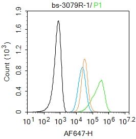
Blank control: Raw264.7.
Primary Antibody (green line): Rabbit Anti-phospho-MCSF Receptor (Tyr923) antibody (SL3079R)
Dilution: 1μg /10^6 cells;
Isotype Control Antibody (orange line): Rabbit IgG .
Secondary Antibody : Goat anti-rabbit IgG-AF647
Dilution: 1μg /test.
Protocol
The cells were incubated in 5%BSA to block non-specific protein-protein interactions for 30 min at room temperature .Cells stained with Primary Antibody for 30 min at room temperature. The secondary antibody used for 40 min at room temperature. Acquisition of 20,000 events was performed.
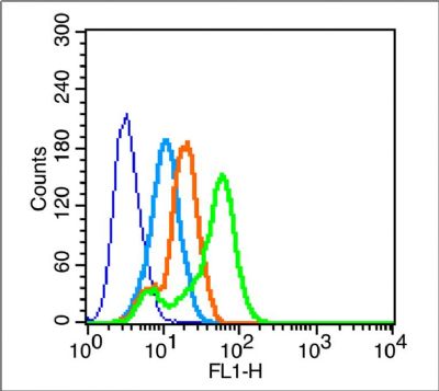
Blank control (blue line): MCF7 (blue).
Primary Antibody (green line): Rabbit Anti-phospho-MCSF Receptor(Tyr923) antibody (SL3079R)
Dilution: 1μg /10^6 cells;
Isotype Control Antibody (orange line): Rabbit IgG .
Secondary Antibody (white blue line): F(ab’)2 fragment goat anti-rabbit IgG-FITC
Dilution: 1μg /test.
Protocol
The cells were fixed with 2% paraformaldehyde for 10 min at room temperature.Cells stained with Primary Antibody for 30 min at room temperature. The cells were then incubated in 1 X PBS/2%BSA/10% goat serum to block non-specific protein-protein interactions followed by the antibody for 15 min at room temperature. The secondary antibody used for 40 min at room temperature. Acquisition of 20,000 events was performed.
Product Feedback Wall
Message :
Your Email :
Copyright © 2007-2018 Sunlong Medical All Rights Reserved.