[IF=13.751] Allison A Fitzgerald. et al. DPP inhibition alters the CXCR3 axis and enhances NK and CD8+ T cell infiltration to improve anti-PD1 efficacy in murine models of pancreatic ductal adenocarcinoma. J Immunother Cancer. 2021 Nov;9(11):e002837 IHC ; Mouse.
[IF=4.044] Malte Puchert. et al. Identification of CXCL11 as part of chemokine network controlling skeletal muscle development. 2021 Jan 27 WB,IF,IHC ; Rat.
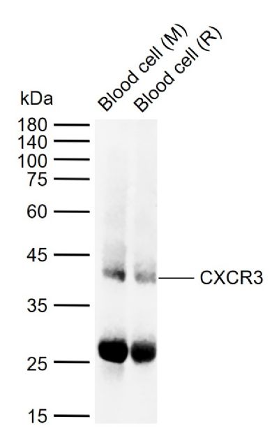
[IF=3.078] Puchert M et al. CXCL11 promotes tumor progression by the biased use of the chemokine receptors CXCR3 and CXCR7. Cytokine. 2019 Aug 19;125:15969. WB ; Human.
[IF=0] Deyhle et al. CXCL10 increases in human skeletal muscle following damage but is not necessary for muscle regeneration. (2018) Physiol.Rep. 6:e13689 IHC ; mouse.
[IF=4.88] Massafra, Vittoria, et al. "Splenic dendritic cell involvement in FXR-mediated amelioration of DSS colitis." Biochimica et Biophysica Acta (BBA)-Molecular Basis of Disease (2015). IHSLCP ; Mouse.
[IF=4.2] Ge, L. P., et al. "Integration of nondegradable polystyrene and degradable gelatin in a core–sheath nanofibrous patch for pelvic reconstruction." International Journal of Nanomedicine 2015:10 3193–641 IHSLCP ; Rat.
[IF=2.784] Yin M et al. Protective effects of CXCR3/HO‑1 gene‑modified BMMSCs on damaged intestinal epithelial cells: Role of the p38‑MAPK signaling pathway.Int J Mol Med. 2019 May;43(5):2086-2102. ICC ; Mouse.
[IF=5.51] Baur et al. The Transcription Factor NFATc1 Supports the Rejection of Heterotopic Heart Allografts. (2018) Front.Immunol. 9:1338 IHC ; Mouse.
[IF=2] Wang, Jun, et al. "Anti‐inflammatory and retinal protective effects of capsaicin on ischemia‐induced injuries through the release of endogenous somatostatin." Clinical and Experimental Pharmacology and Physiology (2017). IHSLCP ; Mouse.
[IF=3.06] Zhang, Wen, et al. "The role of CXCR3 in the induction of primary biliary cirrhosis." Clinical and Developmental Immunology 2011 (2011). IHSLCP ; Mouse.
[IF=5.94] Saxena, Amit, et al. "CXCR3-independent actions of the CXC chemokine CXCL10 in the infarcted myocardium and in isolated cardiac fibroblasts are mediated through proteoglycans." Cardiovascular Research (2014): cvu138. Mouse.