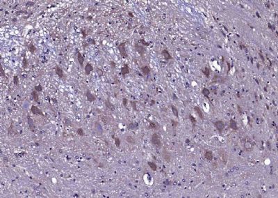
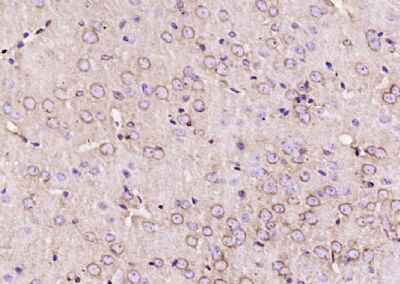
Paraformaldehyde-fixed, paraffin embedded (Rat cerebellum); Antigen retrieval by boiling in sodium citrate buffer (pH6.0) for 15min; Block endogenous peroxidase by 3% hydrogen peroxide for 20 minutes; Blocking buffer (normal goat serum) at 37°C for 30min; Antibody incubation with (CACNA1G) Polyclonal Antibody, Unconjugated (SL2781R) at 1:200 overnight at 4°C, followed by operating according to SP Kit(Rabbit) (sp-0023) instructionsand DAB staining
Paraformaldehyde-fixed, paraffin embedded (mouse brain); Antigen retrieval by boiling in sodium citrate buffer (pH6.0) for 15min; Block endogenous peroxidase by 3% hydrogen peroxide for 20 minutes; Blocking buffer (normal goat serum) at 37°C for 30min; Antibody incubation with (CACNA1G) Polyclonal Antibody, Unconjugated (SL2781R) at 1:200 overnight at 4°C, followed by operating according to SP Kit(Rabbit) (sp-0023) instructionsand DAB staining.
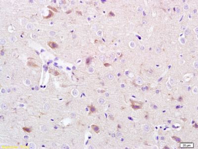
Tissue/cell: rat brain tissue; 4% Paraformaldehyde-fixed and paraffin-embedded;
Antigen retrieval: citrate buffer ( 0.01M, pH 6.0 ), Boiling bathing for 15min; Block endogenous peroxidase by 3% Hydrogen peroxide for 30min; Blocking buffer (normal goat serum,SLC0005) at 37℃ for 20 min;
Incubation: Anti-CACNA1G/Cav3.1 Polyclonal Antibody, Unconjugated(SL2781R) 1:200, overnight at 4°C, followed by conjugation to the secondary antibody(SP-0023) and DAB(SLC0010) staining
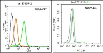
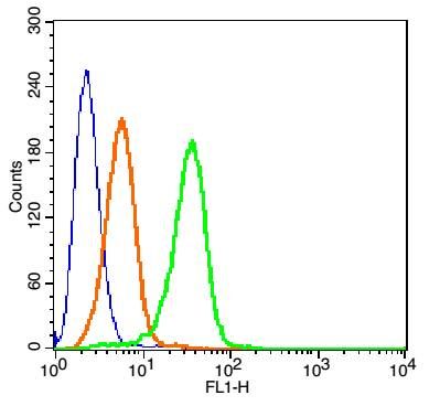
Black line : Positive blank control (MCF7); Negative blank control (431)
Green line : Primary Antibody (Rabbit Anti-CACNA1G antibody (SL2781R) )
Orange line:Isotype Control Antibody (Rabbit IgG) .
Blue line : Secondary Antibody (Goat anti-rabbit IgG-AF488)
MCF7(Positive)and A431(Negative control)cells (black) were incubated in 5% BSA blocking buffer for 30 min at room temperature. Cells were then stained with CACNA1G Antibody(SL2781R)at 1:50 dilution in blocking buffer and incubated for 30 min at room temperature, washed twice with 2% BSA in PBS, followed by secondary antibody(blue) incubation for 40 min at room temperature. Acquisitions of 20,000 events were performed. Cells stained with primary antibody (green), and isotype control (orange).
Blank control(blue): MCF7 Cells(fixed with 2% paraformaldehyde (10 min). The cells were washed twice with 1 X PBS).
Primary Antibody: Rabbit Anti- CACNA1G/AF488 Conjugated antibody (SL2781R-AF488), Dilution: 1μg in 100 μL 1X PBS containing 0.5% BSA;
Isotype Control Antibody: Rabbit IgG/AF488(orange) ,used under the same conditions.
|