Home > Product > Antibody > Rabbit Anti-RIPK3 antibody
Receptor interacting protein 3; Receptor interacting serine threonine kinase 3; Receptor interacting serine/threonine protein kinase 3; Receptor-interacting protein 3; Receptor-interacting serine/threonine-protein kinase 3; RIP 3; RIP like protein kinase
Cat:
SL3551R
Species Reactivity:
Human,Mouse,Rat,(predicted: Dog,Pig,Cow,Rabbit,Guinea Pig,)
Immunogen:
KLH conjugated synthetic peptide derived from human RIPK3:101-230/518
Format:
Liquid
Storage instructions:
Shipped at 4℃. Store at -20 °C for one year. Avoid repeated freeze/thaw cycles.
Concentration:
1mg/ml
Clonality:
Polyclonal
Isotype:
IgG
Applications:
WB=1:500-2000ELISA=1:5000-10000IHC-P=1:100-500IHC-F=1:100-500IF=1:100-500(Paraffin sections need to do antigen repair)not yet tested in other applications.optimal dilutions/concentrations should be determined by the end user.
Host:
Rabbit
Product Overview:
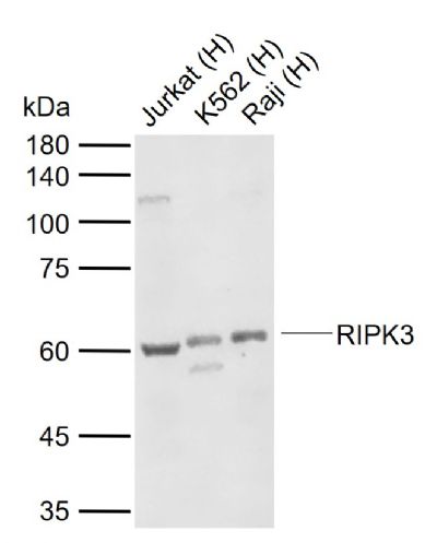
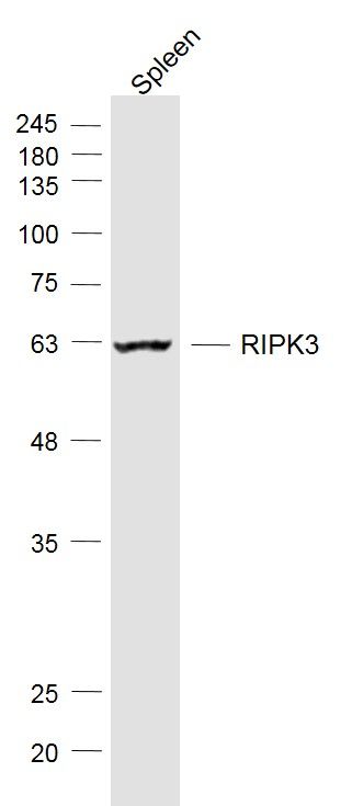
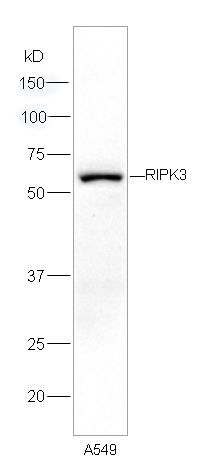
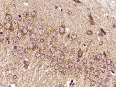
Sample: Lane 1: Human Jurkat cell lysates Lane 2: Human K562 cell lysates Lane 3: Human Raji cell lysates Primary: Anti-RIPK3 (SL3551R) at 1/1000 dilutionSecondary: IRDye800CW Goat Anti-Rabbit IgG at 1/20000 dilutionPredicted band size: 57 kDaObserved band size: 60 kDaSample: Spleen (Mouse) Lysate at 40 ugPrimary: Anti-RIPK3 (SL3551R) at 1/1000 dilutionSecondary: IRDye800CW Goat Anti-Rabbit IgG at 1/20000 dilutionPredicted band size: 57 kDObserved band size: 57 kDSample: A549 cells lysates, 30ug; Primary: Anti-RIPK3 (SL3551R) at 1:300 dilution; Secondary: HRP conjugated Goat Anti-Rabbit IgG(SL0295G-HRP) at 1: 5000 dilution; Predicted band size : 57kDObserved band size : 57kDParaformaldehyde-fixed, paraffin embedded (Rat brain); Antigen retrieval by boiling in sodium citrate buffer (pH6.0) for 15min; Block endogenous peroxidase by 3% hydrogen peroxide for 20 minutes; Blocking buffer (normal goat serum) at 37°C for 30min; Antibody incubation with (RIPK3) Polyclonal Antibody, Unconjugated (SL3551R) at 1:400 overnight at 4°C, followed by a conjugated secondary antibody (sp-0023) for 20 minutes and DAB staining.Tissue/cell: human pneumonia tissue; 4% Paraformaldehyde-fixed and paraffin-embedded; Antigen retrieval: citrate buffer ( 0.01M, pH 6.0 ), Boiling bathing for 15min; Block endogenous peroxidase by 3% Hydrogen peroxide for 30min; Blocking buffer (normal goat serum,SLC0005) at 37℃ for 20 min; Incubation: Anti-RIPK3 Polyclonal Antibody, Unconjugated(SL3551R) 1:200, overnight at 4°C, followed by conjugation to the secondary antibody(SP-0023) and DAB(SLC0010) staining
More
Unit:
Price: $
Product PDFs
Datasheet:


The product of this gene is a member of the receptor-interacting protein (RIP) family of serine/threonine protein kinases, and contains a SLCterminal domain unique from other RIP family members. The encoded protein is predominantly localized to the cytoplasm, and can undergo nucleocytoplasmic shuttling dependent on novel nuclear localization and export signals. It is a component of the tumor necrosis factor (TNF) receptor-I signaling complex, and can induce apoptosis and weakly activate the NF-kappaB transcription factor. [provided by RefSeq, Jul 2008]

Function:
Essential for programmed necrosis in response to death-inducing TNF-alpha family members. Upon induction of necrosis, RIPK3 interacts with, and phosphorylates RIPK1 to form a necrosis-inducing complex. RIPK3 binds to and enhances the activity of three metabolic enzymes: GLUL, GLUD1, and PYGL. These metabolic enzymes may eventually stimulate the tricarboxylic acid cycle and oxidative phosphorylation, which could result in enhanced ROS production.

Subunit:
Interacts (via RIP homotypic interaction motif) with RIPK1 (via RIP homotypic interaction motif); this interaction induces RIPK1 phosphorylation and formation of a RIPK1-RIPK3 necrosis-inducing complex. Upon TNF-induced necrosis, the RIPK1-RIPK3 dimer further interacts with PGAM5 and MLKL; the formation of this complex leads to PGAM5 phosphorylation and increase in PGAM5 phosphatase activity. Binds TRAF2 and is recruited to the TNFR-1 signaling complex. Interacts with PYGL, GLUL and GLUD1; these interactions result in activation of these metabolic enzymes. Interacts with BIRC2/c-IAP1, BIRC3/c-IAP2 and XIAP/BIRC4.

Subcellular Location:
Cytoplasm. Cell membrane. Mitochondrion (Potential).

Tissue Specificity:
Highly expressed in the pancreas. Detected at lower levels in heart, placenta, lung and kidney. Isoform 3 is significantly increased in colon and lung cancers.

Post-translational modifications:
RIPK1 and RIPK3 undergo reciprocal auto- and trans-phosphorylation. Phosphorylation of Ser-199 plays a role in the necroptotic function of RIPK3.
Polyubiquitinated with 'Lys-48' and 'Lys-63'-linked chains by BIRC2/c-IAP1 and BIRC3/c-IAP2, leading to activation of NF-kappa-B.

Similarity:
Belongs to the protein kinase superfamily. TKL Ser/Thr protein kinase family.
Contains 1 protein kinase domain.

SWISS:
Q9Y572

Gene ID:
11035

Database links:

Entrez Gene: 11035 Human

Entrez Gene: 56532 Mouse

Omim: 605817 Human

SwissProt: Q9Y572 Human

SwissProt: Q9QZL0 Mouse

Unigene: 268551 Human

Unigene: 46612 Mouse


Picture

Sample:
Lane 1: Human Jurkat cell lysates
Lane 2: Human K562 cell lysates
Lane 3: Human Raji cell lysates
Primary: Anti-RIPK3 (SL3551R) at 1/1000 dilution
Secondary: IRDye800CW Goat Anti-Rabbit IgG at 1/20000 dilution
Predicted band size: 57 kDa
Observed band size: 60 kDa
Sample:
Spleen (Mouse) Lysate at 40 ug
Primary: Anti-RIPK3 (SL3551R) at 1/1000 dilution
Secondary: IRDye800CW Goat Anti-Rabbit IgG at 1/20000 dilution
Predicted band size: 57 kD
Observed band size: 57 kD
Sample: A549 cells lysates, 30ug;
Primary: Anti-RIPK3 (SL3551R) at 1:300 dilution;
Secondary: HRP conjugated Goat Anti-Rabbit IgG(SL0295G-HRP) at 1: 5000 dilution;
Predicted band size : 57kD
Observed band size : 57kD
Paraformaldehyde-fixed, paraffin embedded (Rat brain); Antigen retrieval by boiling in sodium citrate buffer (pH6.0) for 15min; Block endogenous peroxidase by 3% hydrogen peroxide for 20 minutes; Blocking buffer (normal goat serum) at 37°C for 30min; Antibody incubation with (RIPK3) Polyclonal Antibody, Unconjugated (SL3551R) at 1:400 overnight at 4°C, followed by a conjugated secondary antibody (sp-0023) for 20 minutes and DAB staining.
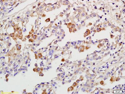
Tissue/cell: human pneumonia tissue; 4% Paraformaldehyde-fixed and paraffin-embedded;
Antigen retrieval: citrate buffer ( 0.01M, pH 6.0 ), Boiling bathing for 15min; Block endogenous peroxidase by 3% Hydrogen peroxide for 30min; Blocking buffer (normal goat serum,SLC0005) at 37℃ for 20 min;
Incubation: Anti-RIPK3 Polyclonal Antibody, Unconjugated(SL3551R) 1:200, overnight at 4°C, followed by conjugation to the secondary antibody(SP-0023) and DAB(SLC0010) staining
Product Feedback Wall
Message :
Your Email :
Copyright © 2007-2018 Sunlong Medical All Rights Reserved.