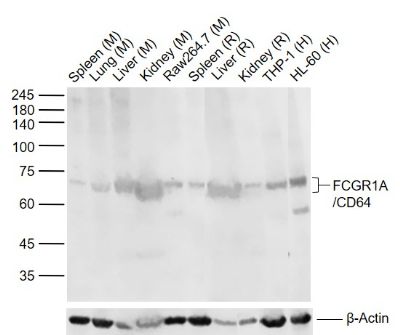
Sample:
Lane 1: Mouse Spleen tissue lysates
Lane 2: Mouse Lung tissue lysates
Lane 3: Mouse Liver tissue lysates
Lane 4: Mouse Kidney tissue lysates
Lane 5: Mouse Raw264.7 cell lysates
Lane 6: Rat Spleen tissue lysates
Lane 7: Rat Liver tissue lysates
Lane 8: Rat Kidney tissue lysates
Lane 9: Human THP-1 cell lysates
Lane 10: Human HL60 cell lysates
Primary: Anti- FCGR1A/CD64 (SL3511R) at 1/1000 dilution
Secondary: IRDye800CW Goat Anti-Rabbit IgG at 1/20000 dilution
Predicted band size: 41 kDa
Observed band size: 70 kDa
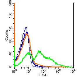
Blank control: Mouse spleen cells(blue).
Primary Antibody: Rabbit Anti- FCGR1A/CD64 antibody(SL3511R), Dilution: 5μg in 100 μL 1X PBS containing 0.5% BSA;
Isotype Control Antibody: Rabbit IgG (orange) ,used under the same conditions.
Secondary Antibody: Goat anti-rabbit IgG-PE(white blue), Dilution: 1:200 in 1 X PBS containing 0.5% BSA.
Protocol
Primary antibody (SL3511R, 5μg /1x10^6 cells) were incubated for 30 min on the ice, followed by 1 X PBS containing 0.5% BSA + 1 0% goat serum (15 min) to block non-specific protein-protein interactions. Then the Goat Anti-rabbit IgG/PE antibody was added into the blocking buffer mentioned above to react with the primary antibody at 1/200 dilution for 30 min on ice. Acquisition of 20,000 events was performed.
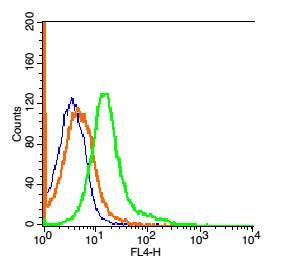
Blank control: 293T Cells(blue).
Primary Antibody: Rabbit Anti-FCGR1A/CD64/AF647 Conjugated antibody (SL3511R-AF647), Dilution: 1μg in 100 μL 1X PBS containing 0.5% BSA;
Isotype Control Antibody: Rabbit IgG/AF647(orange) ,used under the same conditions.
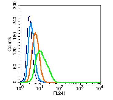
Blank control(blue): U937(fixed with 2% paraformaldehyde (10 min)).
Primary Antibody: Rabbit Anti-CD64 antibody(SL3511R), Dilution: 1μg in 100 μL 1X PBS containing 0.5% BSA;
Isotype Control Antibody: Rabbit IgG(orange) ,used under the same conditions );
Secondary Antibody: Goat anti-rabbit IgG-PE(white blue), Dilution: 1:200 in 1 X PBS containing 0.5% BSA.
|