Perilipins, members of the PAT protein family (named after lipid droplet proteins Perilipin, Adipophilin, and TIP47) are found exclusively at the surface of lipid droplets in adipocytes and steroidogenic cells. They have been suggested to function as regulators of lipolysis and triacylglycerol storage within adipose tissue. Four distinct isoforms ranging from perilipin A (57 kDa) to perilipin D (26 kDa) have been identified and they share an identical amino terminal sequences, and contain 2–6 consensus protein kinase A (PKA) phosphorylation sites. Perilipin C and D have been detected only in steroidogenic cells. Perilipin A is the most abundant form on the lipid droplets of adipocytes. The phosphorylation of perilipin by PKA, which is accompanied by the phosphorylation and translocation of hormone-sensitive lipase from the cytosol to the lipid droplets, promotes lipolysis. There is evidence for the presence of perilipin A in atheroma plaques suggesting that the protein may be involved in the development of therosclerosis by controlling as in adipocytes the hydrolysis of stored lipids.
Function:
Modulator of adipocyte lipid metabolism. Coats lipid storage droplets to protect them from breakdown by hormone-sensitive lipase (HSL). Its absence may result in leanness.
Subunit:
Interacts with ASHD5.
Subcellular Location:
Lipid droplet. Note=Lipid droplet surface-associated.
Tissue Specificity:
Adipocytes.
Post-translational modifications:
Major cAMP-dependent protein kinase-substrate in adipocytes, also dephosphorylated by PP1. When phosphorylated, may be maximally sensitive to HSL and when unphosphorylated, may play a role in the inhibition of lipolysis, by acting as a barrier in lipid droplet.
DISEASE:
Defects in PLIN1 are the cause of familial partial lipodystrophy type 4 (FPLD4) [MIM:613877]. FPLD4 is a form of lipodystrophy characterized by loss of subcutaneous adipose tissue primarily affecting the lower limbs, insulin-resistant diabetes mellitus, hypertriglyceridemia, and hypertension.
Similarity:
Belongs to the perilipin family.
SWISS:
O6048
Gene ID:
5346
Database links:
Entrez Gene: 520598 Cow
Entrez Gene: 5346 Human
Entrez Gene: 103968 Mouse
Entrez Gene: 25629 Rat
Omim: 170290 Human
SwissProt: A4IFB3 Cow
SwissProt: O6048 Human
SwissProt: Q8CGN5 Mouse
SwissProt: P43884 Rat
Unigene: 103253 Human
Unigene: 254917 Mouse
Unigene: 9737 Rat
脂肪细胞中贮存的甘油三酯的水解是人和动物体内精细调节的动态平衡过程,与维持机体能量的动态平衡和代谢正常密切相关。Perilipin A是成熟脂肪细胞中脂滴膜上表达量较多的一种结构和功能蛋白,是多条脂解通路的终末靶点之一,对脂肪细胞的脂肪分解起着关键的调控作用.
perilipin可能在脂肪分解调控中起到“分子开关”的作用。蛋白激酶A(PKA)、细胞外信号调节激酶(ERK)等信号转导通路参与了脂肪分解。肿瘤坏死因子仅(TNFα)、过氧化物酶体增殖物激活受体γ(PPAγ)激动剂、瘦素(leptin)均可以影响perilipin的表达。近年来研究,perilipin可通过蛋白酶体途径来调节其蛋白量的表达。脂肪分解调控中的关键蛋白perilipin对2型糖尿病、肥胖、动脉粥样硬化等多种代谢性疾病及心血管疾病相关联。
| Picture |
Sample: Thymus (Mouse) Lysate at 40 ug
Primary: Anti-Perilipin A (SL3789R) at 1/300 dilution
Secondary: IRDye800CW Goat Anti-Rabbit IgG at 1/20000 dilution
Predicted band size: 57 kD
Observed band size: 57 kD
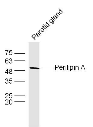
Sample:Parotid gland (Mouse) Lysate at 40 ug
Primary: Anti-Perilipin A (SL3789R) at 1/300 dilution
Secondary: IRDye800CW Goat Anti-Rabbit IgG at 1/20000 dilution
Predicted band size: 57 kD
Observed band size: 57 kD
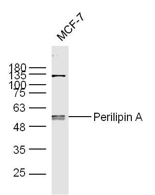
Sample:
MCF-7 Cell (Human) Lysate at 30 ug
Primary: Anti-Perilipin A (Bs- 3789R) at 1/300 dilution
Secondary: IRDye800CW Goat Anti-Rabbit IgG at 1/20000 dilution
Predicted band size: 57 kD
Observed band size: 57 kD
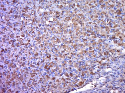
Paraformaldehyde-fixed, paraffin embedded (rat ovary); Antigen retrieval by boiling in sodium citrate buffer (pH6.0) for 15min; Block endogenous peroxidase by 3% hydrogen peroxide for 20 minutes; Blocking buffer (normal goat serum) at 37°C for 30min; Antibody incubation with (Perilipin A ) Polyclonal Antibody, Unconjugated (SL3789R) at 1:400 overnight at 4°C, followed by a conjugated secondary antibody (sp-0023) for 20 minutes and DAB staining.
|
|
|