Home > Product > Antibody > Rabbit Anti-phospho-Alpha synuclein (Ser129) antibody
SNCA(phospho S129); alpha Synuclein (phospho S129) ;alpha-Synuclein (Phospho-Ser129); Synuclein alpha(Phospho-Ser129); SNCA; SYUA_HUMAN; Alpha synuclein; Alpha-synuclein, isoform NACP140; alpha SYN; MGC105443; MGC110988; MGC127560; MGC64356; NACP; Non A b
Cat:
SL5628R
Species Reactivity:
Human,Mouse,Rat,(predicted: Dog,Pig,Cow,Horse,Rabbit,Sheep,)
Immunogen:
KLH conjugated Synthesised phosphopeptide derived from human SNCA around the phosphorylation site of Ser129:MP(p-S)EE
Format:
Liquid
Storage instructions:
Shipped at 4℃. Store at -20 °C for one year. Avoid repeated freeze/thaw cycles.
Concentration:
1mg/ml
Clonality:
Polyclonal
Isotype:
IgG
Applications:
WB=1:500-2000ELISA=1:5000-10000IHC-P=1:100-500IHC-F=1:100-500Flow-Cyt=1ug/TestIF=1:100-500(Paraffin sections need to do antigen repair)not yet tested in other applications.optimal dilutions/concentrations should be determined by the end user.
Host:
Rabbit
Product Overview:
Sample: RAW264.7 (Mouse) CellLysate at 40 ugPrimary: Anti-phospho-Alpha synuclein(Ser129)(SL5628R) at 1/300 dilutionSecondary: IRDye800CW Goat Anti-Rabbit IgG at 1/20000 dilutionPredicted band size: 15 kDObserved band size: 15 kDSample: Lane 1: Human SY5Y cell lysates Lane 2: Human U87MG cell lysates Lane 3: Human U251 cell lysates Lane 4: Human HUVEC Cell Lysates Lane 5: Human HELA Cell Lysates Lane 6: Human 293T Cell Lysates Primary: Anti-phospho-Alpha synuclein (Ser129) (SL5628R) at 1/1000 dilutionSecondary: IRDye800CW Goat Anti-Rabbit IgG at 1/20000 dilutionPredicted band size: 15 kDaObserved band size: 18 kDaSample: Lane 1: Cerebellum (Mouse) Lysate at 40 ugLane 2: Cerebrum (Mouse) Lysate at 40 ugLane 3: Cerebellum (Rat) Lysate at 40 ugLane 4: Cerebrum (Rat) Lysate at 40 ugLane 5: SH-SY5Y (Human) Cell Lysate at 30 ugLane 6: U251 (Human) Cell Lysate at 30 ugLane 7: 293T (Human) Cell Lysate at 30 ugLane 8: Hela (Human) Cell Lysate at 30 ugPrimary: Anti-phospho-Alpha synuclein (Ser129) (SL5628R) at 1/1000 dilutionSecondary: IRDye800CW Goat Anti-Rabbit IgG at 1/20000 dilutionPredicted band size: 18 kDObserved band size: 18 kDBlank control (Black line): HUVEC (Black). Primary Antibody (green line): Rabbit Anti-phospho-Alpha synuclein(Ser129) antibody (SL5628R) Dilution: 3μg /10^6 cells; Isotype Control Antibody (orange line): Rabbit IgG .Secondary Antibody (white blue line): Goat anti-rabbit IgG-AF647Dilution: 1μg /test. ProtocolThe cells were fixed with 4% PFA (10min at room temperature)and then permeabilized with 90% ice-cold methanol for 20 min at room temperature. The cells were then incubated in 5%BSA to block non-specific protein-protein interactions for 30 min at room temperature .Cells stained with Primary Antibody for 30 min at room temperature. The secondary antibody used for 40 min at room temperature. Acquisition of 20,000 events was performed.
More
Unit:
Price: $
Product PDFs
Datasheet:


Alpha-synuclein is a member of the synuclein family, which also includes beta- and gamma-synuclein. Synucleins are abundantly expressed in the brain and alpha- and beta-synuclein inhibit phospholipase D2 selectively. SNCA may serve to integrate presynaptic signaling and membrane trafficking. Defects in SNCA have been implicated in the pathogenesis of Parkinson disease. SNCA peptides are a major component of amyloid plaques in the brains of patients with Alzheimer's disease. Alternatively spliced transcripts encoding different isoforms have been identified for this gene. [provided by RefSeq, Feb 2016]..

Function:
May be involved in the regulation of dopamine release and transport. Induces fibrillization of microtubule-associated protein tau. Reduces neuronal responsiveness to various apoptotic stimuli, leading to a decreased caspase-3 activation.

Subunit:
Soluble monomer which can form filamentous aggregates. Interacts with UCHL1. Interacts with phospholipase D and histones.

Subcellular Location:
Cytoplasm. Membrane. Nucleus. Cell junction, synapse. Note=Membrane-bound in dopaminergic neurons.

Tissue Specificity:
Expressed principally in brain but is also expressed in low concentrations in all tissues examined except in liver. Concentrated in presynaptic nerve terminals.

Post-translational modifications:
Note=Genetic alterations of SNCA resulting in aberrant polymerization into fibrils, are associated with several neurodegenerative diseases (synucleinopathies). SNCA fibrillar aggregates represent the major non A-beta component of Alzheimer disease amyloid plaque, and a major component of Lewy body inclusions. They are also found within Lewy body (LB)-like intraneuronal inclusions, glial inclusions and axonal spheroids in neurodegeneration with brain iron accumulation type 1.

Defects in SNCA are the cause of Parkinson disease type 1 (PARK1) [MIM:168601]. A complex neurodegenerative disorder characterized by bradykinesia, resting tremor, muscular rigidity and postural instability. Additional features are characteristic postural abnormalities, dysautonomia, dystonic cramps, and dementia. The pathology of Parkinson disease involves the loss of dopaminergic neurons in the substantia nigra and the presence of Lewy bodies (intraneuronal accumulations of aggregated proteins), in surviving neurons in various areas of the brain. The disease is progressive and usually manifests after the age of 50 years, although early-onset cases (before 50 years) are known. The majority of the cases are sporadic suggesting a multifactorial etiology based on environmental and genetic factors. However, some patients present with a positive family history for the disease. Familial forms of the disease usually begin at earlier ages and are associated with atypical clinical features.

Defects in SNCA are the cause of Parkinson disease type 4 (PARK4) [MIM:605543]. A complex neurodegenerative disorder with manifestations ranging from typical Parkinson disease to dementia with Lewy bodies. Clinical features include parkinsonian symptoms (tremor, rigidity, postural instability and bradykinesia), dementia, diffuse Lewy body pathology, autonomic dysfunction, hallucinations and paranoia.

Defects in SNCA are the cause of dementia Lewy body (DLB) [MIM:127750]. A neurodegenerative disorder clinically characterized by mental impairment leading to dementia, parkinsonism, often with fluctuating cognitive function, visual hallucinations, falls, syncopal episodes, and sensitivity to neuroleptic medication. Brainstem or cortical intraneuronal accumulations of aggregated proteins (Lewy bodies) are the only essential pathologic features. Patients may also have hippocampal and neocortical senile plaques, sometimes in sufficient number to fulfill the diagnostic criteria for Alzheimer disease.

DISEASE:
Note=Genetic alterations of SNCA resulting in aberrant polymerization into fibrils, are associated with several neurodegenerative diseases (synucleinopathies). SNCA fibrillar aggregates represent the major non A-beta component of Alzheimer disease amyloid plaque, and a major component of Lewy body inclusions. They are also found within Lewy body (LB)-like intraneuronal inclusions, glial inclusions and axonal spheroids in neurodegeneration with brain iron accumulation type 1.

Defects in SNCA are the cause of Parkinson disease type 1 (PARK1) [MIM:168601]. A complex neurodegenerative disorder characterized by bradykinesia, resting tremor, muscular rigidity and postural instability. Additional features are characteristic postural abnormalities, dysautonomia, dystonic cramps, and dementia. The pathology of Parkinson disease involves the loss of dopaminergic neurons in the substantia nigra and the presence of Lewy bodies (intraneuronal accumulations of aggregated proteins), in surviving neurons in various areas of the brain. The disease is progressive and usually manifests after the age of 50 years, although early-onset cases (before 50 years) are known. The majority of the cases are sporadic suggesting a multifactorial etiology based on environmental and genetic factors. However, some patients present with a positive family history for the disease. Familial forms of the disease usually begin at earlier ages and are associated with atypical clinical features.

Defects in SNCA are the cause of Parkinson disease type 4 (PARK4) [MIM:605543]. A complex neurodegenerative disorder with manifestations ranging from typical Parkinson disease to dementia with Lewy bodies. Clinical features include parkinsonian symptoms (tremor, rigidity, postural instability and bradykinesia), dementia, diffuse Lewy body pathology, autonomic dysfunction, hallucinations and paranoia.

Defects in SNCA are the cause of dementia Lewy body (DLB) [MIM:127750]. A neurodegenerative disorder clinically characterized by mental impairment leading to dementia, parkinsonism, often with fluctuating cognitive function, visual hallucinations, falls, syncopal episodes, and sensitivity to neuroleptic medication. Brainstem or cortical intraneuronal accumulations of aggregated proteins (Lewy bodies) are the only essential pathologic features. Patients may also have hippocampal and neocortical senile plaques, sometimes in sufficient number to fulfill the diagnostic criteria for Alzheimer disease.

Similarity:
Belongs to the synuclein family.

SWISS:
P37168

Gene ID:
6622

Database links:

Entrez Gene: 6622 Human

Entrez Gene: 20617 Mouse

Entrez Gene: 29219 Rat

Omim: 163890 Human

SwissProt: P37168 Human

SwissProt: O55042 Mouse

SwissProt: P37377 Rat

Unigene: 21374 Human

Unigene: 17484 Mouse

Unigene: 1827 Rat


Synuclein 包括α-Synuclein,β-Synuclein 和γ-Synuclein 是神经细胞中富含的前突触蛋白。α-Synuclein,Alzheimer’(AD)病淀粉样蛋白沉积的成份之一,集中分布在神经细胞的包体和突触。在帕金森病人中发现有α-Synuclein的变异型,而γ-Synuclein与轴突病理学有关。此抗体将为Lewy小体痴呆症、Parkinson症、AD和其它一些神经性疾病提供有用的病理诊断。 Picture

Sample:
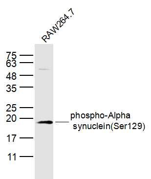
RAW264.7 (Mouse) CellLysate at 40 ug
Primary: Anti-phospho-Alpha synuclein(Ser129)(SL5628R) at 1/300 dilution
Secondary: IRDye800CW Goat Anti-Rabbit IgG at 1/20000 dilution
Predicted band size: 15 kD
Observed band size: 15 kD
Sample:
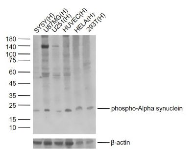
Lane 1: Human SY5Y cell lysates
Lane 2: Human U87MG cell lysates
Lane 3: Human U251 cell lysates
Lane 4: Human HUVEC Cell Lysates
Lane 5: Human HELA Cell Lysates
Lane 6: Human 293T Cell Lysates
Primary: Anti-phospho-Alpha synuclein (Ser129) (SL5628R) at 1/1000 dilution
Secondary: IRDye800CW Goat Anti-Rabbit IgG at 1/20000 dilution
Predicted band size: 15 kDa
Observed band size: 18 kDa
Sample:
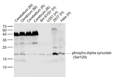
Lane 1: Cerebellum (Mouse) Lysate at 40 ug
Lane 2: Cerebrum (Mouse) Lysate at 40 ug
Lane 3: Cerebellum (Rat) Lysate at 40 ug
Lane 4: Cerebrum (Rat) Lysate at 40 ug
Lane 5: SH-SY5Y (Human) Cell Lysate at 30 ug
Lane 6: U251 (Human) Cell Lysate at 30 ug
Lane 7: 293T (Human) Cell Lysate at 30 ug
Lane 8: Hela (Human) Cell Lysate at 30 ug
Primary: Anti-phospho-Alpha synuclein (Ser129) (SL5628R) at 1/1000 dilution
Secondary: IRDye800CW Goat Anti-Rabbit IgG at 1/20000 dilution
Predicted band size: 18 kD
Observed band size: 18 kD
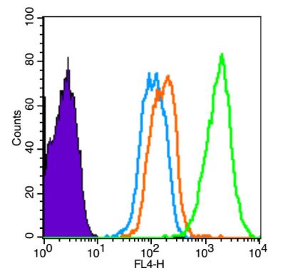
Blank control (Black line): HUVEC (Black). Primary Antibody (green line): Rabbit Anti-phospho-Alpha synuclein(Ser129) antibody (SL5628R) Dilution: 3μg /10^6 cells; Isotype Control Antibody (orange line): Rabbit IgG . Secondary Antibody (white blue line): Goat anti-rabbit IgG-AF647 Dilution: 1μg /test. Protocol The cells were fixed with 4% PFA (10min at room temperature)and then permeabilized with 90% ice-cold methanol for 20 min at room temperature. The cells were then incubated in 5%BSA to block non-specific protein-protein interactions for 30 min at room temperature .Cells stained with Primary Antibody for 30 min at room temperature. The secondary antibody used for 40 min at room temperature. Acquisition of 20,000 events was performed.
Product Feedback Wall
Message :
Your Email :
Copyright © 2007-2018 Sunlong Medical All Rights Reserved.