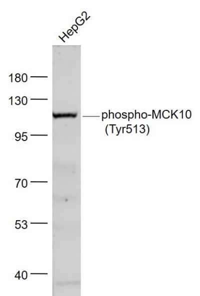
Sample:
HepG2(Human) Cell Lysate at 30 ug
Primary: Anti- phospho-MCK10 (Tyr513) (SL6269R) at 1/1000 dilution
Secondary: IRDye800CW Goat Anti-Rabbit IgG at 1/20000 dilution
Predicted band size: 99 kD
Observed band size: 120 kD
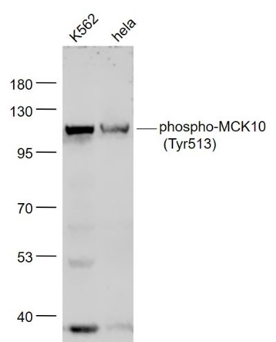
Sample:
K562(Human) Cell Lysate at 30 ug
Hela(Human) Cell Lysate at 30 ug
Primary: Anti- phospho-MCK10 (Tyr513) (SL6269R) at 1/1000 dilution
Secondary: IRDye800CW Goat Anti-Rabbit IgG at 1/20000 dilution
Predicted band size: 99 kD
Observed band size: 120 kD
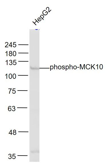
Sample:
HepG2(Human) Cell Lysate at 30 ug
Primary: Anti-phospho-MCK10 (SL6269R) at 1/1000 dilution
Secondary: IRDye800CW Goat Anti-Rabbit IgG at 1/20000 dilution
Predicted band size: 99 kD
Observed band size: 120 kD
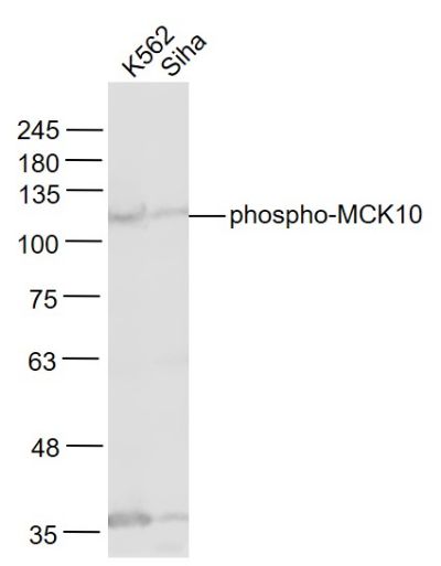
Sample:
K562(Human) Cell Lysate at 30 ug
Siha(Human) Cell Lysate at 30 ug
Primary: Anti-phospho-MCK10 (SL6269R) at 1/1000 dilution
Secondary: IRDye800CW Goat Anti-Rabbit IgG at 1/20000 dilution
Predicted band size: 99 kD
Observed band size: 120 kD
|