Home > Product > Antibody > Rabbit Anti-IMPDH1 antibody
IMDH1_HUMAN; IMP (inosine monophosphate) dehydrogenase 1; IMP dehydrogenase 1; IMPD 1; IMPD; IMPD1; IMPDH 1; IMPDH I; IMPDH-I; Impdh1; Inosine 5' monophosphate dehydrogenase 1; Inosine monophosphate dehydrogenase 1; Inosine-5''-monophosphate dehydrogenase
Cat:
SL6256R
Species Reactivity:
Human,Mouse,Rat,(predicted: Dog,Pig,Cow,Rabbit,Guinea Pig,)
Immunogen:
KLH conjugated synthetic peptide derived from human IMPDH1:201-300/514
Format:
Liquid
Storage instructions:
Shipped at 4℃. Store at -20 °C for one year. Avoid repeated freeze/thaw cycles.
Concentration:
1mg/ml
Clonality:
Polyclonal
Isotype:
IgG
Applications:
WB=1:500-2000ELISA=1:5000-10000IHC-P=1:100-500IHC-F=1:100-500Flow-Cyt=1ug/TestIF=1:100-500(Paraffin sections need to do antigen repair)not yet tested in other applications.optimal dilutions/concentrations should be determined by the end user.
Host:
Rabbit
Product Overview:
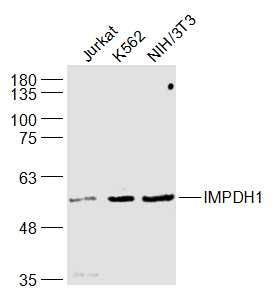
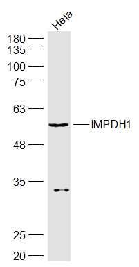
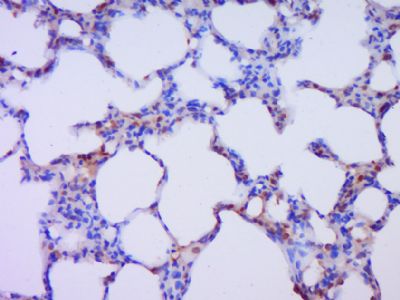
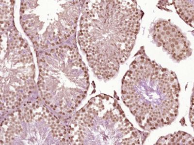
Sample: Jurkat(Human) Cell Lysate at 30 ugK562(Human) cell Lysate at 30 ugNIH/3T3(Mouse) Cell Lysate at 30 ugPrimary: Anti-IMPDH1 (SL6256R) at 1/1000 dilutionSecondary: IRDye800CW Goat Anti-Rabbit IgG at 1/20000 dilutionPredicted band size: 55 kDObserved band size: 55 kDSample: Hela(Human) Cell Lysate at 30 ugPrimary: Anti-IMPDH1 (SL6256R) at 1/1000 dilutionSecondary: IRDye800CW Goat Anti-Rabbit IgG at 1/20000 dilutionPredicted band size: 55 kDObserved band size: 55 kDParaformaldehyde-fixed, paraffin embedded (Rat lung); Antigen retrieval by boiling in sodium citrate buffer (pH6.0) for 15min; Block endogenous peroxidase by 3% hydrogen peroxide for 20 minutes; Blocking buffer (normal goat serum) at 37°C for 30min; Antibody incubation with (IMPDH1) Polyclonal Antibody, Unconjugated (SL6256R) at 1:400 overnight at 4°C, followed by operating according to SP Kit(Rabbit) (sp-0023) instructionsand DAB staining. Paraformaldehyde-fixed, paraffin embedded (Mouse testis); Antigen retrieval by boiling in sodium citrate buffer (pH6.0) for 15min; Block endogenous peroxidase by 3% hydrogen peroxide for 20 minutes; Blocking buffer (normal goat serum) at 37°C for 30min; Antibody incubation with (IMPDH1) Polyclonal Antibody, Unconjugated (SL6256R) at 1:400 overnight at 4°C, followed by operating according to SP Kit(Rabbit) (sp-0023) instructionsand DAB staining.Blank control:Hela. Primary Antibody (green line): Rabbit Anti-IMPDH1 antibody (SL6256R) Dilution: 1ug/Test; Secondary Antibody : Goat anti-rabbit IgG-FITCDilution: 0.5ug/Test. ProtocolThe cells were fixed with 4% PFA (10min at room temperature)and then permeabilized with 0.1% PBST for 20 min at room temperature.The cells were then incubated in 5%BSA to block non-specific protein-protein interactions for 30 min at room temperature .Cells stained with Primary Antibody for 30 min at room temperature. The secondary antibody used for 40 min at room temperature. Acquisition of 20,000 events was performed.
More
Unit:
Price: $
Product PDFs
Datasheet:


The protein encoded by this gene acts as a homotetramer to regulate cell growth. The encoded protein is an enzyme that catalyzes the synthesis of xanthine monophosphate (XMP) from inosine-5'-monophosphate (IMP). This is the rate-limiting step in the de novo synthesis of guanine nucleotides. Defects in this gene are a cause of retinitis pigmentosa type 10 (RP10). Several transcript variants encoding different isoforms have been found for this gene. [provided by RefSeq, Dec 2008].

Function:
Catalyzes the conversion of inosine 5'-phosphate (IMP) to xanthosine 5'-phosphate (XMP), the first committed and rate-limiting step in the de novo synthesis of guanine nucleotides, and therefore plays an important role in the regulation of cell growth. Could also have a single-stranded nucleic acid-binding activity and could play a role in RNA and/or DNA metabolism. It may also have a role in the development of malignancy and the growth progression of some tumors.

Subunit:
Homotetramer.

Subcellular Location:
Cytoplasm. Nucleus.

Tissue Specificity:
IMP type I is the main species in normal leukocytes and type II predominates over type I in the tumor.

DISEASE:
Retinitis pigmentosa 10 (RP10) [MIM:36105]: A retinal dystrophy belonging to the group of pigmentary retinopathies. Retinitis pigmentosa is characterized by retinal pigment deposits visible on fundus examination and primary loss of rod photoreceptor cells followed by secondary loss of cone photoreceptors. Patients typically have night vision blindness and loss of midperipheral visual field. As their condition progresses, they lose their far peripheral visual field and eventually central vision as well. Note=The disease is caused by mutations affecting the gene represented in this entry.
Leber congenital amaurosis 11 (LCA11) [MIM:613837]: A severe dystrophy of the retina, typically becoming evident in the first years of life. Visual function is usually poor and often accompanied by nystagmus, sluggish or near-absent papillary responses, photophobia, high hyperopia and keratoconus. Note=The disease is caused by mutations affecting the gene represented in this entry.

Similarity:
Belongs to the IMPDH/GMPR family.
Contains 2 CBS domains.

SWISS:
P20839

Gene ID:
3614

Database links:

Entrez Gene: 3614 Human

Entrez Gene: 23917 Mouse

Entrez Gene: 362329 Rat

Omim: 146690 Human

SwissProt: P20839 Human

SwissProt: P50096 Mouse

SwissProt: D3ZLZ7 Rat

Unigene: 65881 Human

Unigene: 260707 Mouse

Unigene: 104925 Rat


Picture

Sample:
Jurkat(Human) Cell Lysate at 30 ug
K562(Human) cell Lysate at 30 ug
NIH/3T3(Mouse) Cell Lysate at 30 ug
Primary: Anti-IMPDH1 (SL6256R) at 1/1000 dilution
Secondary: IRDye800CW Goat Anti-Rabbit IgG at 1/20000 dilution
Predicted band size: 55 kD
Observed band size: 55 kD
Sample:
Hela(Human) Cell Lysate at 30 ug
Primary: Anti-IMPDH1 (SL6256R) at 1/1000 dilution
Secondary: IRDye800CW Goat Anti-Rabbit IgG at 1/20000 dilution
Predicted band size: 55 kD
Observed band size: 55 kD
Paraformaldehyde-fixed, paraffin embedded (Rat lung); Antigen retrieval by boiling in sodium citrate buffer (pH6.0) for 15min; Block endogenous peroxidase by 3% hydrogen peroxide for 20 minutes; Blocking buffer (normal goat serum) at 37°C for 30min; Antibody incubation with (IMPDH1) Polyclonal Antibody, Unconjugated (SL6256R) at 1:400 overnight at 4°C, followed by operating according to SP Kit(Rabbit) (sp-0023) instructionsand DAB staining.
Paraformaldehyde-fixed, paraffin embedded (Mouse testis); Antigen retrieval by boiling in sodium citrate buffer (pH6.0) for 15min; Block endogenous peroxidase by 3% hydrogen peroxide for 20 minutes; Blocking buffer (normal goat serum) at 37°C for 30min; Antibody incubation with (IMPDH1) Polyclonal Antibody, Unconjugated (SL6256R) at 1:400 overnight at 4°C, followed by operating according to SP Kit(Rabbit) (sp-0023) instructionsand DAB staining.
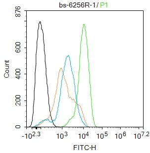
Blank control:Hela.
Primary Antibody (green line): Rabbit Anti-IMPDH1 antibody (SL6256R)
Dilution: 1ug/Test;
Secondary Antibody : Goat anti-rabbit IgG-FITC
Dilution: 0.5ug/Test.
Protocol
The cells were fixed with 4% PFA (10min at room temperature)and then permeabilized with 0.1% PBST for 20 min at room temperature.The cells were then incubated in 5%BSA to block non-specific protein-protein interactions for 30 min at room temperature .Cells stained with Primary Antibody for 30 min at room temperature. The secondary antibody used for 40 min at room temperature. Acquisition of 20,000 events was performed.
Product Feedback Wall
Message :
Your Email :
Copyright © 2007-2018 Sunlong Medical All Rights Reserved.