NGL-1 is a single pass type I membrane protein that acts as a cell adhesion molecule. It contains nine leucine-rich repeats (LRR) and one Ig-like C2-type domain. NGL-1 is predominantly expressed in the striatum and the cerebral cortex of both the embryonic and adult brain. NGL-1 specifically interacts with Netrin G1 (a molecule involved in axon guidance in the developing central nervous system) via its LRR region. NGL-1 plays a role in the regulation of neurite outgrowth of developing thalamic neurons. Soluble NGL-1 inhibits thalamic axon outgrowth while NGL-1 that is bound to the surface of developing thalamocortical axons stimulates growth. NGL-1 also interacts with Whirlin possibly stablizing interstereociliar links.
Function:
May promote neurite outgrowth of developing thalamic neurons.
Subunit:
Interacts with NTNG1 and WHRN.
Subcellular Location:
Membrane.
Tissue Specificity:
Highly expressed in the cerebral cortex, including frontal, parietal and occipital lobes. Putamen, amygdala, hippocampus and medulla oblongata show moderate expression. Caudate nucleus and thalamus express small amounts, whereas other brain regions show very weak or no expression.
Similarity:
Contains 1 Ig-like C2-type (immunoglobulin-like) domain.
Contains 9 LRR (leucine-rich) repeats.
Contains 1 LRRCT domain.
Contains 1 LRRNT domain.
SWISS:
Q9HCJ2
Gene ID:
57689
Database links:
Entrez Gene: 57689 Human
Entrez Gene: 241568 Mouse
Entrez Gene: 311236 Rat
Entrez Gene: 553785 Zebrafish
Omim: 608817 Human
SwissProt: Q9HCJ2 Human
SwissProt: Q8C031 Mouse
Unigene: 745123 Human
Unigene: 241682 Mouse
Unigene: 482999 Mouse
Unigene: 219303 Rat
| Picture |
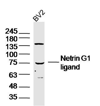
Sample: BV2 Cell (Mouse) Lysate at 40 ug
Primary: Anti- Netrin G1 ligand (SL6710R) at 1/300 dilution
Secondary: IRDye800CW Goat Anti-Rabbit IgG at 1/20000 dilution
Predicted band size: 70 kD
Observed band size: 76 kD
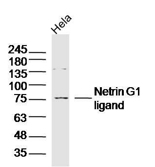
Sample: Hela Cell (Human) Lysate at 40 ug
Primary: Anti- Netrin G1 ligand (SL6710R) at 1/300 dilution
Secondary: IRDye800CW Goat Anti-Rabbit IgG at 1/20000 dilution
Predicted band size: 70 kD
Observed band size: 76 kD
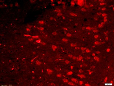
Tissue/cell: mouse brain tissue;4% Paraformaldehyde-fixed and paraffin-embedded;
Antigen retrieval: citrate buffer ( 0.01M, pH 6.0 ), Boiling bathing for 15min; Blocking buffer (normal goat serum,SLC0005) at 37℃ for 20 min;
Incubation: Anti-NGL1 Polyclonal Antibody, Unconjugated(SL6710R) 1:200, overnight at 4°C; The secondary antibody was Goat Anti-Rabbit IgG, Cy3 conjugated(SL0295G-Cy3)used at 1:200 dilution for 40 minutes at 37°C. DAPI(5ug/ml,blue,SLC0033) was used to stain the cell nuclei
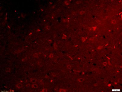
Tissue/cell: rat brain tissue;4% Paraformaldehyde-fixed and paraffin-embedded;
Antigen retrieval: citrate buffer ( 0.01M, pH 6.0 ), Boiling bathing for 15min; Blocking buffer (normal goat serum,SLC0005) at 37℃ for 20 min;
Incubation: Anti-NGL1 Polyclonal Antibody, Unconjugated(SL6710R) 1:200, overnight at 4°C; The secondary antibody was Goat Anti-Rabbit IgG, Cy3 conjugated(SL0295G-Cy3)used at 1:200 dilution for 40 minutes at 37°C. DAPI(5ug/ml,blue,SLC0033) was used to stain the cell nuclei
|
|
|