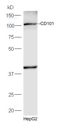
Protein: HepG2(human) lysates at 30ug;
Primary: rabbit Anti-CD101 (SL10727R) at 1:300;
Secondary: HRP conjugated Goat-Anti-rabbit IgG(SL0295G-HRP) at 1: 5000;
Predicted band size:113 kD
Observed band size:113 kD
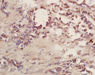
Tissue/cell: human lung carcinoma; 4% Paraformaldehyde-fixed and paraffin-embedded;
Antigen retrieval: citrate buffer ( 0.01M, pH 6.0 ), Boiling bathing for 15min; Block endogenous peroxidase by 3% Hydrogen peroxide for 30min; Blocking buffer (normal goat serum,SLC0005) at 37℃ for 20 min;
Incubation: Anti-CD101 Polyclonal Antibody, Unconjugated(SL10727R) 1:200, overnight at 4°C, followed by conjugation to the secondary antibody(SP-0023) and DAB(SLC0010) staining
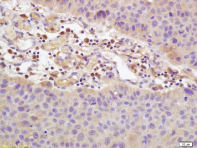
Tissue/cell: human bladder carcinoma; 4% Paraformaldehyde-fixed and paraffin-embedded;
Antigen retrieval: citrate buffer ( 0.01M, pH 6.0 ), Boiling bathing for 15min; Block endogenous peroxidase by 3% Hydrogen peroxide for 30min; Blocking buffer (normal goat serum,SLC0005) at 37℃ for 20 min;
Incubation: Anti-CD101 Polyclonal Antibody, Unconjugated(SL10727R) 1:200, overnight at 4°C, followed by conjugation to the secondary antibody(SP-0023) and DAB(SLC0010) staining
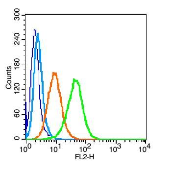
Blank control(blue): U937(fixed with 2% paraformaldehyde (10 min)).
Primary Antibody:Rabbit Anti-CD101 antibody(SL10727R), Dilution: 1μg in 100 μL 1X PBS containing 0.5% BSA;
Isotype Control Antibody: Rabbit IgG(orange) ,used under the same conditions );
Secondary Antibody: Goat anti-rabbit IgG-PE(white blue), Dilution: 1:200 in 1 X PBS containing 0.5% BSA.
|