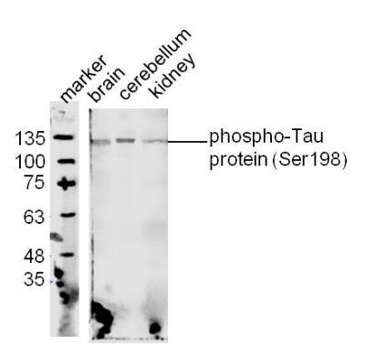
Protein: 1.brain lyates(mo); 2.cerebellum lyates(mo);3.kidney lysates (mo);
Primary: Rabbit Anti-phospho-Tau protein (Ser198) (SL4199R) at 1:300;
Secondary: 800CW Conjugated Goat (polyclonal) Anti-Rabbit IgG(H+L) at 1: 10000;
Predicted band size:79 kD
Observed band size:128 kD
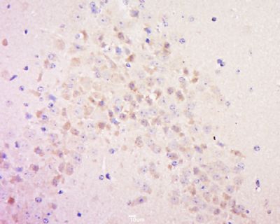
Paraformaldehyde-fixed, paraffin embedded (Rat brain); Antigen retrieval by boiling in sodium citrate buffer (pH6.0) for 15min; Block endogenous peroxidase by 3% hydrogen peroxide for 20 minutes; Blocking buffer (normal goat serum) at 37°C for 30min; Antibody incubation with (phospho-Tau protein (Ser198)) Polyclonal Antibody, Unconjugated (SL4199R) at 1:400 overnight at 4°C, followed by operating according to SP Kit(Rabbit) (sp-0023) instructions and DAB staining.
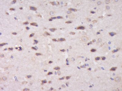
Tissue/cell: Mouse brain tissue; 4% Paraformaldehyde-fixed and paraffin-embedded;
Antigen retrieval: citrate buffer ( 0.01M, pH 6.0 ), Boiling bathing for 15min; Block endogenous peroxidase by 3% Hydrogen peroxide for 30min; Blocking buffer (normal goat serum,SLC0005) at 37℃ for 20 min;
Incubation: Anti-MAPT Polyclonal Antibody, Unconjugated(SL4199R) 1:500, overnight at 4°C, followed by conjugation to the secondary antibody(SP-0023) and DAB(SLC0010) staining
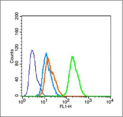
Blank control (blue line): MCF 7 (fixed with 70% methanol (Overnight at 4℃) and then permeabilized with 90% ice-cold methanol for 30 min on ice).
Primary Antibody (green line): Rabbit Anti- phospho-Tau protein (Ser198)antibody (SL4199R),Dilution: 0.2μg /10^5 cells;
Isotype Control Antibody (orange line): Rabbit IgG .
Secondary Antibody (white blue line): Goat anti-rabbit IgG-FITC,Dilution: 1μg /test.
|