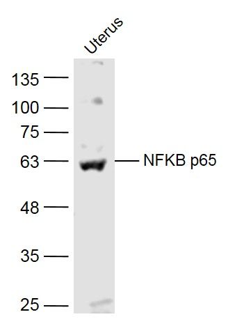
Sample:
Uterus (Mouse) Lysate at 40 ug
Primary: Anti-NFKB p65 (SL20355R) at 1/300 dilution
Secondary: IRDye800CW Goat Anti-Rabbit IgG at 1/20000 dilution
Predicted band size: 62 kD
Observed band size: 62 kD
Paraformaldehyde-fixed, paraffin embedded (rat spleen); Antigen retrieval by boiling in sodium citrate buffer (pH6.0) for 15min; Block endogenous peroxidase by 3% hydrogen peroxide for 20 minutes; Blocking buffer (normal goat serum) at 37°C for 30min; Antibody incubation with (NFKB p65) Polyclonal Antibody, Unconjugated (SL20355R) at 1:200 overnight at 4°C, followed by operating according to SP Kit(Rabbit) (sp-0023) instructionsand DAB staining.
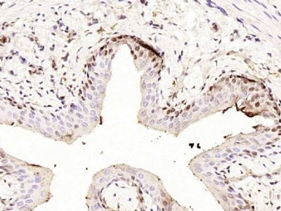
Paraformaldehyde-fixed, paraffin embedded (rat bladder); Antigen retrieval by boiling in sodium citrate buffer (pH6.0) for 15min; Block endogenous peroxidase by 3% hydrogen peroxide for 20 minutes; Blocking buffer (normal goat serum) at 37°C for 30min; Antibody incubation with (NFKB p65) Polyclonal Antibody, Unconjugated (SL20355R) at 1:200 overnight at 4°C, followed by operating according to SP Kit(Rabbit) (sp-0023) instructionsand DAB staining.
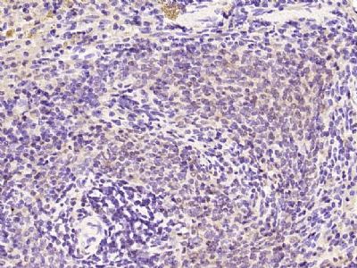
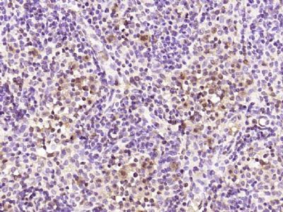
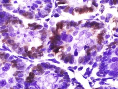
Paraformaldehyde-fixed, paraffin embedded (rat lymphoid); Antigen retrieval by boiling in sodium citrate buffer (pH6.0) for 15min; Block endogenous peroxidase by 3% hydrogen peroxide for 20 minutes; Blocking buffer (normal goat serum) at 37°C for 30min; Antibody incubation with (NFKB p65) Polyclonal Antibody, Unconjugated (SL20355R) at 1:200 overnight at 4°C, followed by operating according to SP Kit(Rabbit) (sp-0023) instructionsand DAB staining.
Paraformaldehyde-fixed, paraffin embedded (Rat colon); Antigen retrieval by boiling in sodium citrate buffer (pH6.0) for 15min; Block endogenous peroxidase by 3% hydrogen peroxide for 20 minutes; Blocking buffer (normal goat serum) at 37°C for 30min; Antibody incubation with (NFKB p65) Polyclonal Antibody, Unconjugated (SL20355R) at 1:400 overnight at 4°C, followed by a conjugated secondary antibody (sp-0023) for 20 minutes and DAB staining.
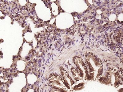
Paraformaldehyde-fixed, paraffin embedded (Mouse small intestine); Antigen retrieval by boiling in sodium citrate buffer (pH6.0) for 15min; Block endogenous peroxidase by 3% hydrogen peroxide for 20 minutes; Blocking buffer (normal goat serum) at 37°C for 30min; Antibody incubation with (NFKB p65 ) Polyclonal Antibody, Unconjugated (SL20355R) at 1:400 overnight at 4°C, followed by a conjugated secondary antibody (sp-0023) for 20 minutes and DAB staining.
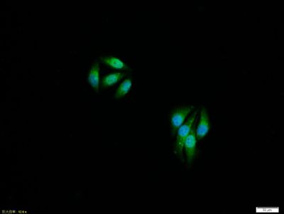
Tissue/cell:MCF7 cell; 4% Paraformaldehyde-fixed; Triton X-100 at room temperature for 20 min; Blocking buffer (normal goat serum,SLC0005) at 37°C for 20 min; Antibody incubation with (NFKB p65) polyclonal Antibody, Unconjugated (SL20355R) 1:100, 90 minutes at 37°C; followed by a FITC conjugated Goat Anti-Rabbit IgG antibody at 37°C for 90 minutes, DAPI (blue, C02-04002) was used to stain the cell nuclei.
Tissue/cell:MCF7 cell; 4% Paraformaldehyde-fixed; Triton X-100 at room temperature for 20 min; Blocking buffer (normal goat serum,SLC0005) at 37°C for 20 min; Antibody incubation with (NFKB p65) polyclonal Antibody, Unconjugated (SL20355R) 1:100, 90 minutes at 37°C; followed by a FITC conjugated Goat Anti-Rabbit IgG antibody at 37°C for 90 minutes, DAPI (blue, C02-04002) was used to stain the cell nuclei.
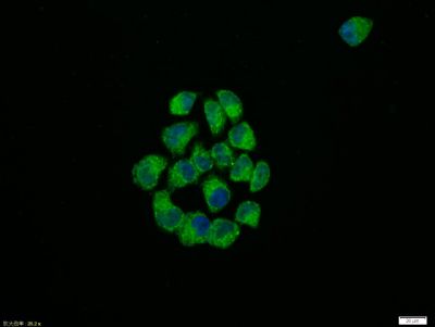
Tissue/cell:Hela cell; 4% Paraformaldehyde-fixed; Triton X-100 at room temperature for 20 min; Blocking buffer (normal goat serum,SLC0005) at 37°C for 20 min; Antibody incubation with (NFKB p65) polyclonal Antibody, Unconjugated (SL20355R) 1:100, 90 minutes at 37°C; followed by a FITC conjugated Goat Anti-Rabbit IgG antibody at 37°C for 90 minutes, DAPI (blue, C02-04002) was used to stain the cell nuclei.
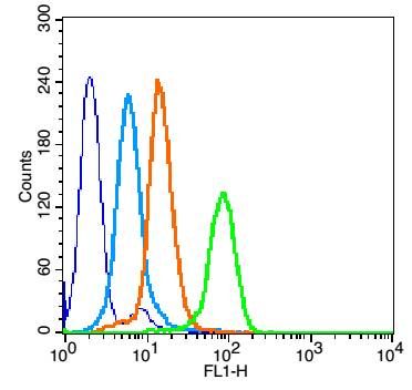
Overlay histogram showing HL 60 cells stained with SL20355R (Green line).
The cells were fixed with 90% methanol (5 min) and then permeabilized with 0.01M PBS-Tween for 20 min. The cells were then incubated in 1x PBS / 10% normal goat serum to block non-specific protein-protein interactions followed by the antibody (SL20355R,1μg/1x10^6 cells) for 30 min at 22℃. The secondary antibody used was fluorescein isothiocyanate goat anti-rabbit IgG (H+L) (SL 0295G-FITC , Brillant blue line) at 1/200 dilution for 30 min at 22℃. Isotype control antibody was rabbit IgG (polyclonal,SL0295P,Orange line) (1μg/1x10^6 cells) used under the same conditions. Unlabelled sample (blue line) was also used as a control. Acquisition of 20,000 events were collected using a 20mW Argon ion laser (488nm) and 525/30 bandpass filter.
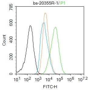
Blank control:A431.
Primary Antibody (green line): Rabbit Anti-NFKB p65 antibody (SL20355R)
Dilution: 1μg /10^6 cells;
Isotype Control Antibody (orange line): Rabbit IgG .
Secondary Antibody : Goat anti-rabbit IgG-FITC
Dilution: 1μg /test.
Protocol
The cells were fixed with 4% PFA (10min at room temperature)and then permeabilized with 90% ice-cold methanol for 20 min at-20℃. The cells were then incubated in 5%BSA to block non-specific protein-protein interactions for 30 min at room temperature .Cells stained with Primary Antibody for 30 min at room temperature. The secondary antibody used for 40 min at room temperature. Acquisition of 20,000 events was performed.
|