Home > Product > Antibody > Rabbit Anti-Caspase-1 P10 antibody
caspase-1 isoform alpha p10 subunit; CASP 1; CASP1; Caspase 1 apoptosis related cysteine peptidase; Caspase 1 apoptosis related cysteine protease; Caspase1; ICE; IL 1 beta converting enzyme; IL 1BC; IL1B convertase; IL1BC; IL1BCE; Interleukin 1 beta conve
Cat:
SL20616R
Species Reactivity:
Human,Mouse,
Immunogen:
KLH conjugated synthetic peptide derived from human Caspase-1 P10:321-404/404
Format:
Liquid
Storage instructions:
Shipped at 4℃. Store at -20 °C for one year. Avoid repeated freeze/thaw cycles.
Concentration:
1mg/ml
Clonality:
Polyclonal
Isotype:
IgG
Applications:
WB=1:500-2000Flow-Cyt=1μg/Testnot yet tested in other applications.optimal dilutions/concentrations should be determined by the end user.
Host:
Rabbit
Product Overview:
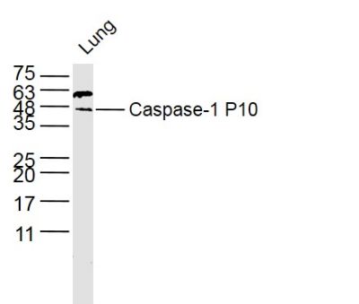
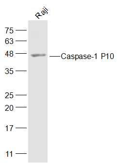
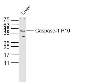
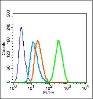
Sample: Lung Cell (Mouse) Lysate at 40 ugPrimary: Anti-NKG2D (SL20616R) at 1/300 dilutionSecondary: IRDye800CW Goat Anti-Rabbit IgG at 1/20000 dilutionPredicted band size: 10/45 kDObserved band size: 45 kDSample: Raji(Human) Cell Lysate at 30 ugPrimary: Anti-Caspase-1 P10 (SL20616R) at 1/300 dilutionSecondary: IRDye800CW Goat Anti-Rabbit IgG at 1/20000 dilutionPredicted band size: 10/45 kDObserved band size: 45 kDSample: Liver Cell (Mouse) Lysate at 40 ugPrimary: Anti-NKG2D (SL20616R) at 1/300 dilutionSecondary: IRDye800CW Goat Anti-Rabbit IgG at 1/20000 dilutionPredicted band size: 10/45 kDObserved band size: 45 kDBlank control (blue line): MCF7(fixed with 70% ethanol (Overnight at 4℃) and then permeabilized with 90% ice-cold methanol for 30 min on ice) Primary Antibody (green line): Rabbit Anti-se-1 P10 antibody (SL20616R),Dilution: 1μg /10^6 cells; Isotype Control Antibody (orange line): Rabbit IgG . Secondary Antibody (white blue line): Goat anti-rabbit IgG-FITC,Dilution: 1μg /test. Blank control:HL-60. Primary Antibody (green line): Rabbit Anti-Caspase-1 P10 antibody (SL20616R) Dilution: 1μg /10^6 cells; Isotype Control Antibody (orange line): Rabbit IgG . Secondary Antibody : Goat anti-rabbit IgG-AF488Dilution: 1μg /test. ProtocolThe cells were fixed with 4% PFA (10min at room temperature)and then permeabilized with 0.1% PBST for 20 min at room temperature. The cells were then incubated in 5%BSA to block non-specific protein-protein interactions for 30 min at room temperature .Cells stained with Primary Antibody for 30 min at room temperature. The secondary antibody used for 40 min at room temperature. Acquisition of 20,000 events was performed.
More
Unit:
Price: $
Product PDFs
Datasheet:


This gene encodes a protein which is a member of the cysteine-aspartic acid protease (caspase) family. Sequential activation of caspases plays a central role in the execution-phase of cell apoptosis. Caspases exist as inactive proenzymes which undergo proteolytic processing at conserved aspartic residues to produce 2 subunits, large and small, that dimerize to form the active enzyme. This gene was identified by its ability to proteolytically cleave and activate the inactive precursor of interleukin-1, a cytokine involved in the processes such as inflammation, septic shock, and wound healing. This gene has been shown to induce cell apoptosis and may function in various developmental stages. Studies of a similar gene in mouse suggest a role in the pathogenesis of Huntington disease. Alternative splicing of this gene results in five transcript variants encoding distinct isoforms. [provided by RefSeq].

Function:
Thiol protease that cleaves IL-1 beta between an Asp and an Ala, releasing the mature cytokine which is involved in a variety of inflammatory processes. Important for defense against pathogens. Cleaves and activates sterol regulatory element binding proteins (SREBPs). Can also promote apoptosis.

Subunit:
Heterotetramer that consists of two anti-parallel arranged heterodimers, each one formed by a 20 kDa (p20) and a 10 kDa (p10) subunit. The p20 subunit can also form a heterodimer with the epsilon isoform which then has an inhibitory effect. May be a component of the inflammasome, a protein complex which also includes PYCARD, CARD8 and NALP2 and whose function would be the activation of proinflammatory caspases. Interacts with CARD17/INCA and CARD18.

Subcellular Location:
Cytoplasm.

Tissue Specificity:
Expressed in larger amounts in spleen and lung. Detected in liver, heart, small intestine, colon, thymus, prostate, skeletal muscle, peripheral blood leukocytes, kidney and testis. No expression in the brain.

Post-translational modifications:
The two subunits are derived from the precursor sequence by an autocatalytic mechanism.

Similarity:
Belongs to the peptidase C14A family.
Contains 1 CARD domain.

SWISS:
P29466

Gene ID:
834

Database links:

Entrez Gene: 834 Human

Entrez Gene: 12362 Mouse

Entrez Gene: 25166 Rat

Omim: 147678 Human

SwissProt: P29466 Human

SwissProt: P29452 Mouse

SwissProt: P43527 Rat

Unigene: 2490 Human

Unigene: 1051 Mouse

Unigene: 37508 Rat


Caspase-1是半胱氨酸蛋白中的一种酶,参与细胞凋亡的调控。它主要用于各种良、恶性肿瘤的研究。 Picture

Sample:
Lung Cell (Mouse) Lysate at 40 ug
Primary: Anti-NKG2D (SL20616R) at 1/300 dilution
Secondary: IRDye800CW Goat Anti-Rabbit IgG at 1/20000 dilution
Predicted band size: 10/45 kD
Observed band size: 45 kD
Sample:
Raji(Human) Cell Lysate at 30 ug
Primary: Anti-Caspase-1 P10 (SL20616R) at 1/300 dilution
Secondary: IRDye800CW Goat Anti-Rabbit IgG at 1/20000 dilution
Predicted band size: 10/45 kD
Observed band size: 45 kD
Sample:
Liver Cell (Mouse) Lysate at 40 ug
Primary: Anti-NKG2D (SL20616R) at 1/300 dilution
Secondary: IRDye800CW Goat Anti-Rabbit IgG at 1/20000 dilution
Predicted band size: 10/45 kD
Observed band size: 45 kD
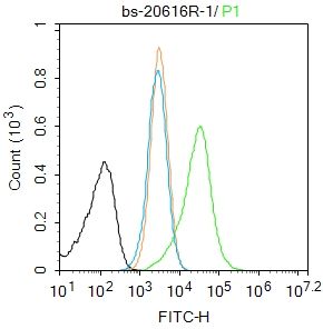
Blank control (blue line): MCF7(fixed with 70% ethanol (Overnight at 4℃) and then permeabilized with 90% ice-cold methanol for 30 min on ice)
Primary Antibody (green line): Rabbit Anti-se-1 P10 antibody (SL20616R),Dilution: 1μg /10^6 cells;
Isotype Control Antibody (orange line): Rabbit IgG .
Secondary Antibody (white blue line): Goat anti-rabbit IgG-FITC,Dilution: 1μg /test.
Blank control:HL-60.
Primary Antibody (green line): Rabbit Anti-Caspase-1 P10 antibody (SL20616R)
Dilution: 1μg /10^6 cells;
Isotype Control Antibody (orange line): Rabbit IgG .
Secondary Antibody : Goat anti-rabbit IgG-AF488
Dilution: 1μg /test.
Protocol
The cells were fixed with 4% PFA (10min at room temperature)and then permeabilized with 0.1% PBST for 20 min at room temperature. The cells were then incubated in 5%BSA to block non-specific protein-protein interactions for 30 min at room temperature .Cells stained with Primary Antibody for 30 min at room temperature. The secondary antibody used for 40 min at room temperature. Acquisition of 20,000 events was performed.
Product Feedback Wall
Message :
Your Email :
Copyright © 2007-2018 Sunlong Medical All Rights Reserved.