Specific References (6) | SL1253R has been referenced in 6 publications.
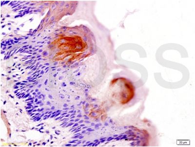
[IF=2.826] Takanori Watanabe et al. Aquaporin 3 Expression in Endometrioid Carcinoma of the Uterine Body Correlated With Early Stage and Lower Grade. Pathol Oncol Res. 2020 Oct;26(4):2247-2253. IHC ; Human.
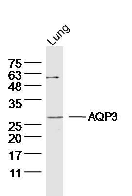
[IF=4.331] Tang SC et al. Glycolic acid attenuates UVB-induced aquaporin-3, matrix metalloproteinase-9 expression, and collagen degradation in keratinocytes and mouse skin. Biochem J. 2019 May 21;476(10):1387-1400. WB ; Human.
[IF=1.935] Sato K et al. Different Prognostic Implications of Aquaporin-1 and Aquaporin-5 Expression among Different Histological Types of Ovarian Carcinoma.Pathol Oncol Res. 2018 Jul 19. IHSLCP ; Human.
[IF=4.24] Zhang, Haifeng, et al. "The AQP-3 water channel and the ClSLC3 chloride channel coordinate the hypotonicity-induced swelling volume in nasopharyngeal carcinoma cells." The International Journal of Biochemistry & Cell Biology (2014). IP ; Human.
[IF=3.414] Li X et al. Physicochemical properties and laxative effects of polysaccharides from Anemarrhena asphodeloides Bge. In loperamide-induced rats. J Ethnopharmacol. 2019 Aug 10;48:111961. WB ; Rat.
[IF=2.311] Haifa Alkhalifa. et al. Inhibition of aquaporins as a potential adjunct to breast cancer cryotherapy. Oncol Lett. 2021 Jun;21(6):1-11 WB ; Human.