Home > Product > Antibody > Rabbit Anti-APAF1(NT) antibody
APAF 1; APAF1; APAF-1; Apoptotic peptidase activating factor 1; Apoptotic protease activating factor; CED 4; CED4; KIAA0413; APAF_HUMAN; Apoptotic protease-activating factor 1.
Cat:
SL0059R
Species Reactivity:
Human,Mouse,Rat,(predicted: Chicken,Dog,Cow,Horse,)
Immunogen:
KLH conjugated synthetic peptide derived from human Apaf-1:13-80/1248
Format:
Liquid
Storage instructions:
Shipped at 4℃. Store at -20 °C for one year. Avoid repeated freeze/thaw cycles.
Concentration:
1mg/ml
Clonality:
Polyclonal
Isotype:
IgG
Applications:
WB=1:500-2000ELISA=1:5000-10000IHC-P=1:100-500IHC-F=1:100-500Flow-Cyt=0.2ug/testICC=1:100IF=1:100-500(Paraffin sections need to do antigen repair)not yet tested in other applications.optimal dilutions/concentrations should be determined by the end user.
Host:
Rabbit
Product Overview:
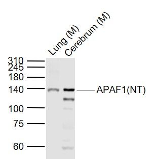
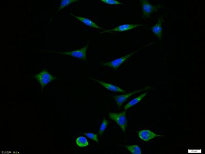
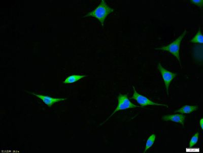
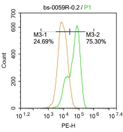
Sample: Lane 1: Lung (Mouse) Lysate at 40 ugLane 2: Cerebrum (Mouse) Lysate at 40 ugPrimary: Anti-APAF1(NT) (SL0059R) at 1/1000 dilutionSecondary: IRDye800CW Goat Anti-Rabbit IgG at 1/20000 dilutionPredicted band size: 140 kDObserved band size: 140 kDParaformaldehyde-fixed, paraffin embedded (rat brain); Antigen retrieval by boiling in sodium citrate buffer (pH6.0) for 15min; Block endogenous peroxidase by 3% hydrogen peroxide for 20 minutes; Blocking buffer (normal goat serum) at 37°C for 30min; Antibody incubation with (APAF1(NT)) Polyclonal Antibody, Unconjugated (SL0059R) at 1:200 overnight at 4°C, followed by operating according to SP Kit(Rabbit) (sp-0023) instructionsand DAB staining.Tissue/cell: rat kidney tissue; 4% Paraformaldehyde-fixed and paraffin-embedded; Antigen retrieval: citrate buffer ( 0.01M, pH 6.0 ), Boiling bathing for 15min; Block endogenous peroxidase by 3% Hydrogen peroxide for 30min; Blocking buffer (normal goat serum,SLC0005) at 37℃ for 20 min; Incubation: Anti-APAF1(NT) Polyclonal Antibody, Unconjugated(SL0059R) 1:200, overnight at 4°C, followed by conjugation to the secondary antibody(SP-0023) and DAB(SLC0010) stainingTissue/cell: rat lung tissue; 4% Paraformaldehyde-fixed and paraffin-embedded; Antigen retrieval: citrate buffer ( 0.01M, pH 6.0 ), Boiling bathing for 15min; Block endogenous peroxidase by 3% Hydrogen peroxide for 30min; Blocking buffer (normal goat serum,SLC0005) at 37℃ for 20 min; Incubation: Anti-APAF1(NT) Polyclonal Antibody, Unconjugated(SL0059R) 1:200, overnight at 4°C, followed by conjugation to the secondary antibody(SP-0023) and DAB(SLC0010) stainingTissue/cell:SH-SY5Y cell; 4% Paraformaldehyde-fixed; Triton X-100 at room temperature for 20 min; Blocking buffer (normal goat serum,SLC0005) at 37°C for 20 min; Antibody incubation with (APAF1(NT)) polyclonal Antibody, Unconjugated (SL0059R) 1:100, 90 minutes at 37°C; followed by a FITC conjugated Goat Anti-Rabbit IgG antibody at 37°C for 90 minutes, DAPI (blue, C02-04002) was used to stain the cell nuclei.Tissue/cell:SH-SY5Y cell; 4% Paraformaldehyde-fixed; Triton X-100 at room temperature for 20 min; Blocking buffer (normal goat serum,SLC0005) at 37°C for 20 min; Antibody incubation with (APAF1(NT)) polyclonal Antibody, Unconjugated (SL0059R) 1:100, 90 minutes at 37°C; followed by a FITC conjugated Goat Anti-Rabbit IgG antibody at 37°C for 90 minutes, DAPI (blue, C02-04002) was used to stain the cell nuclei.U-937 cells were fixed with 4% PFA for 10min at room temperature,permeabilized with 20% PBST for 20 min at room temperature, and incubated in 5% BSA blocking buffer for 30 min at room temperature. Cells were then stained with APAF1(NT) Antibody(SL0059R)at 1:500 dilution in blocking buffer and incubated for 30 min at room temperature, washed twice with 2%BSA in PBS, followed by secondary antibody incubation for 40 min at room temperature. Acquisitions of 20,000 events were performed. Cells stained with primary antibody (green), and isotype control (orange).
More
Unit:
Price: $
Product PDFs
Datasheet:


This gene encodes a cytoplasmic protein that initiates apoptosis. This protein contains several copies of the WD-40 domain, a caspase recruitment domain (CARD), and an ATPase domain(NB-ARC). Upon binding cytochrome c and dATP, this protein forms an oligomeric apoptosome. The apoptosome binds and cleaves caspase 9 preproprotein, releasing its mature, activated form. Activated caspase 9 stimulates the subsequent caspase cascade that commits the cell to apoptosis. Alternative splicing results in several transcript variants encoding different isoforms. [provided by RefSeq, Jul 2008].

Function:
Oligomeric Apaf-1 mediates the cytochrome c-dependent autocatalytic activation of pro-caspase-9 (Apaf-3), leading to the activation of caspase-3 and apoptosis. This activation requires ATP. Isoform 6 is less effective in inducing apoptosis.

Subunit:
Monomer. Oligomerizes upon binding of cytochrome c and dATP. Oligomeric Apaf-1 and pro-caspase-9 bind to each other via their respective NH2-terminal CARD domains and consecutively mature caspase-9 is released from the complex. Pro-caspase-3 is recruited into the Apaf-1-pro-caspase-9 complex via interaction with pro-caspase-9. Interacts with APIP. Interacts (via CARD and NACHT domains) with NAIP/BIRC1 (via NACHT domain).

Subcellular Location:
Cytoplasm.

Tissue Specificity:
Ubiquitous. Highest levels of expression in adult spleen and peripheral blood leukocytes, and in fetal brain, kidney and lung. Isoform 1 is expressed in heart, kidney and liver.

Similarity:
Contains 1 CARD domain.
Contains 1 NB-ARC domain.
Contains 13 WD repeats.

SWISS:
O14727

Gene ID:
317

Database links:

Entrez Gene: 317 Human

Entrez Gene: 11783 Mouse

Entrez Gene: 78963 Rat

Omim: 602233 Human

SwissProt: O14727 Human

SwissProt: O88879 Mouse

SwissProt: Q9EPV5 Rat

Unigene: 728891 Human

Unigene: 220289 Mouse

Unigene: 64522 Rat


Apaf-1 (Apoptosis protease activating factor-1)调节细胞色素C依赖的Caspase-9原的自动催化活性,导致Caspase-3 的激活和引起凋亡。
Apaf-1在成年人的脾脏、外周血白细胞、肾脏、肺和胎儿脑、肾、肺中高水平表达。 Picture

Sample:
Lane 1: Lung (Mouse) Lysate at 40 ug
Lane 2: Cerebrum (Mouse) Lysate at 40 ug
Primary: Anti-APAF1(NT) (SL0059R) at 1/1000 dilution
Secondary: IRDye800CW Goat Anti-Rabbit IgG at 1/20000 dilution
Predicted band size: 140 kD
Observed band size: 140 kD
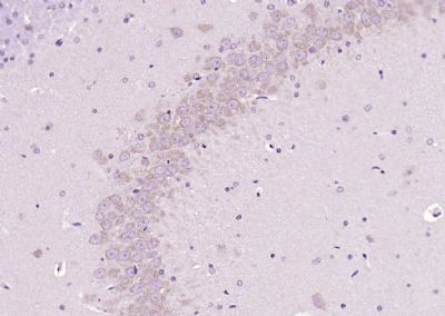
Paraformaldehyde-fixed, paraffin embedded (rat brain); Antigen retrieval by boiling in sodium citrate buffer (pH6.0) for 15min; Block endogenous peroxidase by 3% hydrogen peroxide for 20 minutes; Blocking buffer (normal goat serum) at 37°C for 30min; Antibody incubation with (APAF1(NT)) Polyclonal Antibody, Unconjugated (SL0059R) at 1:200 overnight at 4°C, followed by operating according to SP Kit(Rabbit) (sp-0023) instructionsand DAB staining.
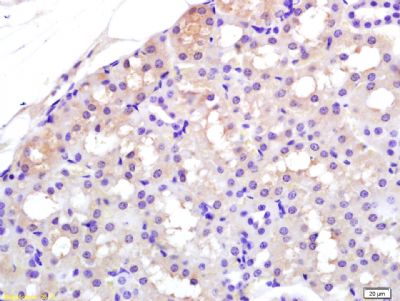
Tissue/cell: rat kidney tissue; 4% Paraformaldehyde-fixed and paraffin-embedded;
Antigen retrieval: citrate buffer ( 0.01M, pH 6.0 ), Boiling bathing for 15min; Block endogenous peroxidase by 3% Hydrogen peroxide for 30min; Blocking buffer (normal goat serum,SLC0005) at 37℃ for 20 min;
Incubation: Anti-APAF1(NT) Polyclonal Antibody, Unconjugated(SL0059R) 1:200, overnight at 4°C, followed by conjugation to the secondary antibody(SP-0023) and DAB(SLC0010) staining
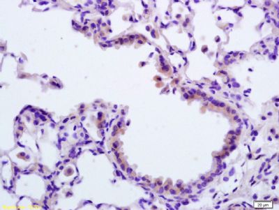
Tissue/cell: rat lung tissue; 4% Paraformaldehyde-fixed and paraffin-embedded;
Antigen retrieval: citrate buffer ( 0.01M, pH 6.0 ), Boiling bathing for 15min; Block endogenous peroxidase by 3% Hydrogen peroxide for 30min; Blocking buffer (normal goat serum,SLC0005) at 37℃ for 20 min;
Incubation: Anti-APAF1(NT) Polyclonal Antibody, Unconjugated(SL0059R) 1:200, overnight at 4°C, followed by conjugation to the secondary antibody(SP-0023) and DAB(SLC0010) staining
Tissue/cell:SH-SY5Y cell; 4% Paraformaldehyde-fixed; Triton X-100 at room temperature for 20 min; Blocking buffer (normal goat serum,SLC0005) at 37°C for 20 min; Antibody incubation with (APAF1(NT)) polyclonal Antibody, Unconjugated (SL0059R) 1:100, 90 minutes at 37°C; followed by a FITC conjugated Goat Anti-Rabbit IgG antibody at 37°C for 90 minutes, DAPI (blue, C02-04002) was used to stain the cell nuclei.
U-937 cells were fixed with 4% PFA for 10min at room temperature,permeabilized with 20% PBST for 20 min at room temperature, and incubated in 5% BSA blocking buffer for 30 min at room temperature. Cells were then stained with APAF1(NT) Antibody(SL0059R)at 1:500 dilution in blocking buffer and incubated for 30 min at room temperature, washed twice with 2%BSA in PBS, followed by secondary antibody incubation for 40 min at room temperature. Acquisitions of 20,000 events were performed. Cells stained with primary antibody (green), and isotype control (orange).
Product Feedback Wall
Message :
Your Email :
Copyright © 2007-2018 Sunlong Medical All Rights Reserved.