Home > Product > Antibody > Rabbit Anti-BTG2 antibody
BTG2_HUMAN; Protein BTG2; BTG family member 2; NGF-inducible anti-proliferative protein PC3; pheochromacytoma cell-3; nerve growth factor-inducible anti-proliferative; B-cell translocation gene 2; BTG2; TIS21; PC3; TIS21.
Cat:
SL0031R
Species Reactivity:
Human,Mouse,(predicted: Rat,Pig,Rabbit,Guinea Pig,)
Immunogen:
KLH conjugated synthetic peptide derived from human BTG2:31-110/158
Format:
Liquid
Storage instructions:
Shipped at 4℃. Store at -20 °C for one year. Avoid repeated freeze/thaw cycles.
Concentration:
1mg/ml
Clonality:
Polyclonal
Isotype:
IgG
Applications:
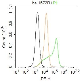
WB=1:500-2000ELISA=1:5000-10000IHC-P=1:100-500IHC-F=1:100-500IF=1:100-500(Paraffin sections need to do antigen repair)not yet tested in other applications.optimal dilutions/concentrations should be determined by the end user.
Host:
Rabbit
Product Overview:
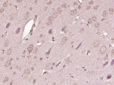
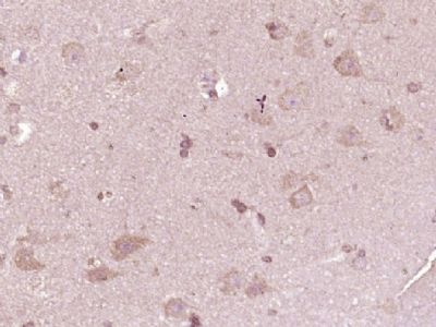
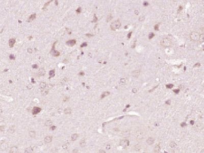
Sample: HepG2 (human)cell Lysate at 40 ugHcclm3 (human)cell Lysate at 40 ugPrimary: Anti- BTG2 (SL0031R)at 1/300 dilutionSecondary: IRDye800CW Goat Anti-Rabbit IgG at 1/20000 dilutionPredicted band size: 17kDObserved band size: 17 kDSample: Lane 1: K562 (Human) Cell Lysate at 30 ugLane 2: 293T (Human) Cell Lysate at 30 ugLane 3: HepG2 (Human) Cell Lysate at 30 ugPrimary: Anti-BTG2 (SL0031R) at 1/1000 dilutionSecondary: IRDye800CW Goat Anti-Rabbit IgG at 1/20000 dilutionPredicted band size: 17 kDObserved band size: 16 kDTissue/cell: mouse heart tissue; 4% Paraformaldehyde-fixed and paraffin-embedded; Antigen retrieval: citrate buffer ( 0.01M, pH 6.0 ), Boiling bathing for 15min; Block endogenous peroxidase by 3% Hydrogen peroxide for 30min; Blocking buffer (normal goat serum,SLC0005) at 37℃ for 20 min; Incubation: Anti-BTG2 Polyclonal Antibody, Unconjugated(SL0031R) 1:200, overnight at 4°C, followed by conjugation to the secondary antibody(SP-0023) and DAB(SLC0010) stainingTissue/cell: mouse small intestine tissue;4% Paraformaldehyde-fixed and paraffin-embedded; Antigen retrieval: citrate buffer ( 0.01M, pH 6.0 ), Boiling bathing for 15min; Blocking buffer (normal goat serum,SLC0005) at 37℃ for 20 min; Incubation: Anti-BTG2 Polyclonal Antibody, Unconjugated(SL0031R) 1:200, overnight at 4°C; The secondary antibody was Goat Anti-Rabbit IgG, Cy3 conjugated(SL0295G-Cy3)used at 1:200 dilution for 40 minutes at 37°C. DAPI(5ug/ml,blue,SLC0033) was used to stain the cell nuclei
More
Unit:
Price: $
Product PDFs
Datasheet:


BTG2 is a member of the BTG/Tob family. This family has structurally related proteins that appear to have antiproliferative properties. BTG2 is involved in the regulation of the G1/S transition of the cell cycle. It modulates transcription regulation mediated by ESR1(referenced from Entrez Gene). BTG2 expression is induced in vivo during neurogenesis, and the gene is transiently expressed in vitro in rat pheochromocytoma PC12 cells after induction of neuronal differentiation by addition of nerve growth factor (NGF); suggesting that BTG2 is functionally significant during the neuronal differentiation process (PMID: 1272398).

Function:
Anti-proliferative protein; the function is mediated by association with deadenylase subunits of the CCR4-NOT complex. Activates mRNA deadenyltion in a CNOT6 and CNOT7-dependent manner. In vitro can inhibit deadenylase activity of CNOT7 and CNOT8. Involved in cell cycle regulation. Could be involved in the growth arrest and differentiation of the neuronal precursors. Modulates transcription regulation mediated by ESR1. Involved in mitochondrial depolarization and neurite outgrowth.

Subunit:
Interacts with PRKCABP. Interacts with CNOT7 and CNOT8; indicative for an asscociation with the CCR4-NOT complex. Interacts with PIN1, inducing mitochondrial depolarization.

Post-translational modifications:
Phosphorylated at Ser-147 by MAPK1/ERK2 and MAPK3/ERK1, and at Ser-149 by MAPK14, leading to PIN1-binding and mitochondrial depolarization.

Similarity:
Belongs to the BTG family.

SWISS:
P78543

Gene ID:
7832

Database links:

Entrez Gene: 7832 Human

Omim: 601597 Human

SwissProt: P78543 Human

Unigene: 519162 Human


Picture

Sample:
HepG2 (human)cell Lysate at 40 ug
Hcclm3 (human)cell Lysate at 40 ug
Primary: Anti- BTG2 (SL0031R)at 1/300 dilution
Secondary: IRDye800CW Goat Anti-Rabbit IgG at 1/20000 dilution
Predicted band size: 17kD
Observed band size: 17 kD
Sample:
Lane 1: K562 (Human) Cell Lysate at 30 ug
Lane 2: 293T (Human) Cell Lysate at 30 ug
Lane 3: HepG2 (Human) Cell Lysate at 30 ug
Primary: Anti-BTG2 (SL0031R) at 1/1000 dilution
Secondary: IRDye800CW Goat Anti-Rabbit IgG at 1/20000 dilution
Predicted band size: 17 kD
Observed band size: 16 kD
Tissue/cell: mouse heart tissue; 4% Paraformaldehyde-fixed and paraffin-embedded;
Antigen retrieval: citrate buffer ( 0.01M, pH 6.0 ), Boiling bathing for 15min; Block endogenous peroxidase by 3% Hydrogen peroxide for 30min; Blocking buffer (normal goat serum,SLC0005) at 37℃ for 20 min;
Incubation: Anti-BTG2 Polyclonal Antibody, Unconjugated(SL0031R) 1:200, overnight at 4°C, followed by conjugation to the secondary antibody(SP-0023) and DAB(SLC0010) staining
Tissue/cell: mouse small intestine tissue;4% Paraformaldehyde-fixed and paraffin-embedded;
Antigen retrieval: citrate buffer ( 0.01M, pH 6.0 ), Boiling bathing for 15min; Blocking buffer (normal goat serum,SLC0005) at 37℃ for 20 min;
Incubation: Anti-BTG2 Polyclonal Antibody, Unconjugated(SL0031R) 1:200, overnight at 4°C; The secondary antibody was Goat Anti-Rabbit IgG, Cy3 conjugated(SL0295G-Cy3)used at 1:200 dilution for 40 minutes at 37°C. DAPI(5ug/ml,blue,SLC0033) was used to stain the cell nuclei
Product Feedback Wall
Message :
Your Email :
Copyright © 2007-2018 Sunlong Medical All Rights Reserved.