Home > Product > Antibody > Rabbit Anti-CD20 antibody
CD20_HUMAN; B-lymphocyte antigen CD20; B-lymphocyte surface antigen B1; Bp35; Leukocyte surface antigen Leu-16; Membrane-spanning 4-domains subfamily A member 1; MS4A1; MS4A2; B1; CVID5; LEU-16; S7.
Cat:
SL0080R
Species Reactivity:
Human,Mouse,Rat,(predicted: Dog,Cow,Rabbit,)
Immunogen:
KLH conjugated synthetic peptide derived from human CD20:133-182/297<Cytoplasmic>
Format:
Liquid
Storage instructions:
Shipped at 4℃. Store at -20 °C for one year. Avoid repeated freeze/thaw cycles.
Concentration:
1mg/ml
Clonality:
Polyclonal
Isotype:
IgG
Applications:
WB=1:500-2000ELISA=1:5000-10000Flow-Cyt=1μg /testnot yet tested in other applications.optimal dilutions/concentrations should be determined by the end user.
Host:
Rabbit
Product Overview:
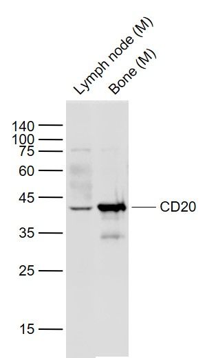
Sample: Lane 1: Lymph node (Mouse) Lysate at 40 ugLane 2: Bone (Mouse) Lysate at 40 ugPrimary: Anti-CD20 (SL0080R) at 1/1000 dilutionSecondary: IRDye800CW Goat Anti-Rabbit IgG at 1/20000 dilutionPredicted band size: 43 kDObserved band size: 43 kDBlank control: U937 (blue). Primary Antibody:Rabbit Anti- CD20 antibody(SL0080R), Dilution: 1μg in 100 μL 1X PBS containing 0.5% BSA; Isotype Control Antibody: Rabbit IgG(orange) ,used under the same conditions ); Secondary Antibody: Goat anti-rabbit IgG-PE(white blue), Dilution: 1:200 in 1 X PBS containing 0.5% BSA.ProtocolThe cells were fixed with 2% paraformaldehyde (10 min) . Primary antibody (SL0080R, 1μg /1x10^6 cells) were incubated for 30 min on the ice, followed by 1 X PBS containing 0.5% BSA + 1 0% goat serum (15 min) to block non-specific protein-protein interactions. Then the Goat Anti-rabbit IgG/PE antibody was added into the blocking buffer mentioned above to react with the primary antibody at 1/200 dilution for 30 min on ice. Acquisition of 20,000 events was performed.
More
Unit:
Price: $
Product PDFs
Datasheet:


CD20 is a non glycosylated protein with a molecular weight of 35 or 37 kDa depending on the degree of phosphorylation. Although not a member of the tetraspanin superfamily of cell surface receptors, it crosses the cell membrane four times. The CD20 antigen is present on human pre B lymphocytes and on B lymphocytes at all stages of maturation, except on plasma cells. Low level expression of the CD20 antigen has been detected on normal T lymphocytes. The CD20 molecule is involved in regulation of B cell differentiation, presumably via its reported function as a Ca++ channel subunit.

Function:
This protein may be involved in the regulation of B-cell activation and proliferation

Subcellular Location:
Membrane; Multi-pass membrane protein.

Tissue Specificity:
Expressed on B-cells.

Post-translational modifications:
Phosphorylated. Might be functionally regulated by protein kinase(s).

DISEASE:
Defects in MS4A1 are the cause of immunodeficiency common variable type 5 (CVID5) [MIM:613495]; also called antibody deficiency due to CD20 defect. CVID5 is a primary immunodeficiency characterized by antibody deficiency, hypogammaglobulinemia, recurrent bacterial infections and an inability to mount an antibody response to antigen. The defect results from a failure of B-cell differentiation and impaired secretion of immunoglobulins; the numbers of circulating B-cells is usually in the normal range, but can be low.

Similarity:
Belongs to the MS4A family.

SWISS:
P11836

Gene ID:
931

Database links:

Entrez Gene: 931 Human

Omim: 112210 Human

SwissProt: P11836 Human

Unigene: 712553 Human


[PHARMACEUTICAL] Monoclonal antibodies (mAb) against CD20 are used to treat B-cell non-Hodgkin lymphoma (NHL). These antibodies include Rituximab (Mabthera), Britumomab (Zevalin) and Tositumomab (Bexxar). CD20 engaged by mAb can generate transmembrane signals capable of directly controlling cell growth and triggering cell death in certain tumors. Alternatively, mAb can mediate complement-dependent cytotoxicity.

B淋巴细胞标志物

CD20是B细胞特异性抗原,其表达呈细胞类型一种质膜蛋白质,参与信号传导作用,此抗体识别大多数B细胞上的一种抗原。多用于标记B淋巴细胞及B细胞淋巴瘤。
CD20一般不与T细胞交叉反应,对B细胞淋巴瘤和急慢性淋巴细胞白血病等有较好的细胞特异性,因此在恶性淋巴瘤,尤其是T或B细胞淋巴瘤的分类上,CD20是最常用的B细胞标志。 Picture

Sample:
Lane 1: Lymph node (Mouse) Lysate at 40 ug
Lane 2: Bone (Mouse) Lysate at 40 ug
Primary: Anti-CD20 (SL0080R) at 1/1000 dilution
Secondary: IRDye800CW Goat Anti-Rabbit IgG at 1/20000 dilution
Predicted band size: 43 kD
Observed band size: 43 kD
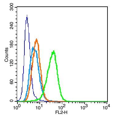
Blank control: U937 (blue).
Primary Antibody:Rabbit Anti- CD20 antibody(SL0080R), Dilution: 1μg in 100 μL 1X PBS containing 0.5% BSA;
Isotype Control Antibody: Rabbit IgG(orange) ,used under the same conditions );
Secondary Antibody: Goat anti-rabbit IgG-PE(white blue), Dilution: 1:200 in 1 X PBS containing 0.5% BSA.
Protocol
The cells were fixed with 2% paraformaldehyde (10 min) . Primary antibody (SL0080R, 1μg /1x10^6 cells) were incubated for 30 min on the ice, followed by 1 X PBS containing 0.5% BSA + 1 0% goat serum (15 min) to block non-specific protein-protein interactions. Then the Goat Anti-rabbit IgG/PE antibody was added into the blocking buffer mentioned above to react with the primary antibody at 1/200 dilution for 30 min on ice. Acquisition of 20,000 events was performed.
Product Feedback Wall
Message :
Your Email :
Copyright © 2007-2018 Sunlong Medical All Rights Reserved.