Home > Product > Antibody > Rabbit Anti-Collagen IV antibody
Arresten; Canstatin; COL4A1; HANAC; ICH; POREN1; Collagen Alpha 1(IV) Chain; Collagen IV Alpha 1 Polypeptide; Collagen Of Basement Membrane Alpha 1 Chain; Collagen Of Basement Membrane Alpha 2 Chain; Collagen Type IV Alpha 1; DKFZp686I14213; FLJ22259; col
Cat:
SL0806R
Species Reactivity:
Human,Rat,Rabbit,(predicted: Mouse,Dog,Pig,Cow,Horse,)
Immunogen:
KLH conjugated synthetic peptide derived from human Collagen alpha-1(IV) chain:1401-1500/1669
Format:
Liquid
Storage instructions:
Shipped at 4℃. Store at -20 °C for one year. Avoid repeated freeze/thaw cycles.
Concentration:
1mg/ml
Clonality:
Polyclonal
Isotype:
IgG
Applications:
ELISA=1:5000-10000IHC-P=1:100-500IHC-F=1:100-500Flow-Cyt=1μg/TestIF=1:100-500(Paraffin sections need to do antigen repair)not yet tested in other applications.optimal dilutions/concentrations should be determined by the end user.
Host:
Rabbit
Product Overview:
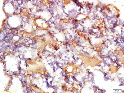
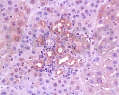
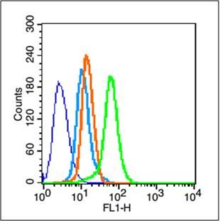
Tissue/cell: rabbit cartilage tissue; 4% Paraformaldehyde-fixed and paraffin-embedded; Antigen retrieval: citrate buffer ( 0.01M, pH 6.0 ), Boiling bathing for 15min; Block endogenous peroxidase by 3% Hydrogen peroxide for 30min; Blocking buffer (normal goat serum,SLC0005) at 37℃ for 20 min; Incubation: Anti-Collagen IV Polyclonal Antibody, Unconjugated(SL0806R) 1:200, overnight at 4°C, followed by conjugation to the secondary antibody(SP-0023) and DAB(SLC0010) stainingTissue/cell: rat liver tissue; 4% Paraformaldehyde-fixed and paraffin-embedded; Antigen retrieval: citrate buffer ( 0.01M, pH 6.0 ), Boiling bathing for 15min; Block endogenous peroxidase by 3% Hydrogen peroxide for 30min; Blocking buffer (normal goat serum,SLC0005) at 37℃ for 20 min; Incubation: Anti-Collagen IV Polyclonal Antibody, Unconjugated(SL0806R) 1:200, overnight at 4°C, followed by conjugation to the secondary antibody(SP-0023) and DAB(SLC0010) stainingBlank control (blue line): Hela (blue). Primary Antibody (green line): Rabbit Anti-Collagen IV antibody (SL0806R) Dilution: 1μg /10^6 cells; Isotype Control Antibody (orange line): Rabbit IgG . Secondary Antibody (white blue line): Goat anti-rabbit IgG-FITCDilution: 1μg /test. ProtocolThe cells were fixed with 70% methanol (Overnight at -20℃) and then permeabilized with ice-cold 90% methanol for 30 min on ice. Cells stained with Primary Antibody for 30 min at room temperature. The cells were then incubated in 1 X PBS/2%BSA/10% goat serum to block non-specific protein-protein interactions followed by the antibody for 15 min at room temperature. The secondary antibody used for 40 min at room temperature. Acquisition of 20,000 events was performed.
More
Unit:
Price: $
Product PDFs
Datasheet:


This gene encodes the major type IV alpha collagen chain of basement membranes. Like the other members of the type IV collagen gene family, this gene is organized in a head-to-head conformation with another type IV collagen gene so that each gene pair shares a common promoter. [provided by RefSeq, Jul 2008]

Function:
Type IV collagen is the major structural component of glomerular basement membranes (GBM), forming a 'chicken-wire' meshwork together with laminins, proteoglycans and entactin/nidogen.
Arresten, comprising the SLCterminal NC1 domain, inhibits angiogenesis and tumor formation. The SLCterminal half is found to possess the anti-angiogenic activity. Specifically inhibits endothelial cell proliferation, migration and tube formation. Inhibits expression of hypoxia-inducible factor 1alpha and ERK1/2 and p38 MAPK activation. Ligand for alpha1/beta1 integrin.

Subunit:
There are six type IV collagen isoforms, alpha 1(IV)-alpha 6(IV), each of which can form a triple helix structure with 2 other chains to generate type IV collagen network.

Subcellular Location:
Secreted, extracellular space, extracellular matrix, basement membrane.

Tissue Specificity:
Highly expressed in placenta.

Post-translational modifications:
Lysines at the third position of the tripeptide repeating unit (G-X-Y) are hydroxylated in all cases and bind carbohydrates.
Prolines at the third position of the tripeptide repeating unit (G-X-Y) are hydroxylated in some or all of the chains.
Type IV collagens contain numerous cysteine residues which are involved in inter- and intramolecular disulfide bonding. 12 of these, located in the NC1 domain, are conserved in all known type IV collagens.
The trimeric structure of the NC1 domains is stabilized by covalent bonds between Lys and Met residues.
Proteolytic processing produces the SLCterminal NC1 peptide, arresten.

DISEASE:
Defects in COL4A1 are a cause of brain small vessel disease with hemorrhage (BSVDH) [MIM:607595]. Brain small vessel diseases underlie 20 to 30 percent of ischemic strokes and a larger proportion of intracerebral hemorrhages. Inheritance is autosomal dominant.
Defects in COL4A1 are the cause of hereditary angiopathy with nephropathy aneurysms and muscle cramps (HANAC) [MIM:611773]. The clinical renal manifestations include hematuria and bilateral large cysts. Histologic analysis revealed complex basement membrane defects in kidney and skin. The systemic angiopathy appears to affect both small vessels and large arteries.
Defects in COL4A1 are a cause of familial porencephaly (POREN1) [MIM:175780]. Porencephaly is a term used for any cavitation or cerebrospinal fluid-filled cyst in the brain. Porencephaly type 1 is usually unilateral and results from focal destructive lesions such as fetal vascular occlusion or birth trauma. Type 2, or schizencephalic porencephaly, is usually symmetric and represents a primary defect or arrest in the development of the cerebral ventricles.

Similarity:
Contains 1 FAD-binding FR-type domain.
Contains 1 ferric oxidoreductase domain.

SWISS:
P02463

Gene ID:
1282

Database links:

Entrez Gene: 1282 Human

Omim: 24070 Human

SwissProt: P02462 Human

Unigene: 17441 Human


IV型胶原为基底膜的主要成分,主要用于各种良恶性组织(乳腺癌、胃肠道癌等)中基底膜分布情况的研究.IV型胶原是构成基底膜的主要成分。该抗体可特异性识别人的IV型胶原,与人类皮肤、肾、肌肉、脾、淋巴结、胎盘和肺的基底膜呈阳性反应,主要用于各种组织癌症中基底膜情况的研究。 Picture

Tissue/cell: rabbit cartilage tissue; 4% Paraformaldehyde-fixed and paraffin-embedded;
Antigen retrieval: citrate buffer ( 0.01M, pH 6.0 ), Boiling bathing for 15min; Block endogenous peroxidase by 3% Hydrogen peroxide for 30min; Blocking buffer (normal goat serum,SLC0005) at 37℃ for 20 min;
Incubation: Anti-Collagen IV Polyclonal Antibody, Unconjugated(SL0806R) 1:200, overnight at 4°C, followed by conjugation to the secondary antibody(SP-0023) and DAB(SLC0010) staining
Tissue/cell: rat liver tissue; 4% Paraformaldehyde-fixed and paraffin-embedded;
Antigen retrieval: citrate buffer ( 0.01M, pH 6.0 ), Boiling bathing for 15min; Block endogenous peroxidase by 3% Hydrogen peroxide for 30min; Blocking buffer (normal goat serum,SLC0005) at 37℃ for 20 min;
Incubation: Anti-Collagen IV Polyclonal Antibody, Unconjugated(SL0806R) 1:200, overnight at 4°C, followed by conjugation to the secondary antibody(SP-0023) and DAB(SLC0010) staining
Blank control (blue line): Hela (blue).
Primary Antibody (green line): Rabbit Anti-Collagen IV antibody (SL0806R)
Dilution: 1μg /10^6 cells;
Isotype Control Antibody (orange line): Rabbit IgG .
Secondary Antibody (white blue line): Goat anti-rabbit IgG-FITC
Dilution: 1μg /test.
Protocol
The cells were fixed with 70% methanol (Overnight at -20℃) and then permeabilized with ice-cold 90% methanol for 30 min on ice. Cells stained with Primary Antibody for 30 min at room temperature. The cells were then incubated in 1 X PBS/2%BSA/10% goat serum to block non-specific protein-protein interactions followed by the antibody for 15 min at room temperature. The secondary antibody used for 40 min at room temperature. Acquisition of 20,000 events was performed.
Product Feedback Wall
Message :
Your Email :
Copyright © 2007-2018 Sunlong Medical All Rights Reserved.