Home > Product > Antibody > Rabbit Anti-ILK-1 antibody
59 kDa serine/threonine protein kinase; 59 kDa serine/threonine-protein kinase; ILK-2; ILK_HUMAN; Integrin linked Kinase; Integrin-linked protein kinase; DKFZp686F1765; EC 2.7.11.1; ILK 1; ILK 2; ILK; ILK1; ILK2; Integrin linked Kinase 2; Integrin linked
Cat:
SL0317R
Species Reactivity:
Human,Mouse,Rat,(predicted: Chicken,Dog,Cow,)
Immunogen:
KLH conjugated synthetic peptide derived from human ILK-1:301-400/452
Format:
Liquid
Storage instructions:
Shipped at 4℃. Store at -20 °C for one year. Avoid repeated freeze/thaw cycles.
Concentration:
1mg/ml
Clonality:
Polyclonal
Isotype:
IgG
Applications:
WB=1:500-2000ELISA=1:5000-10000IHC-P=1:100-500IHC-F=1:100-500IF=1:100-500(Paraffin sections need to do antigen repair)not yet tested in other applications.optimal dilutions/concentrations should be determined by the end user.
Host:
Rabbit
Product Overview:
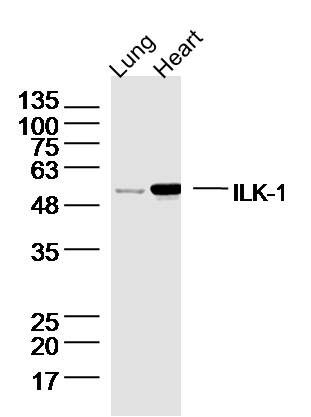
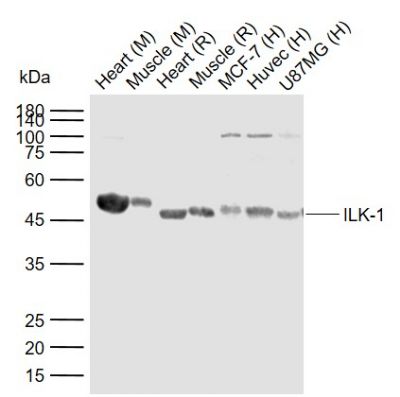
Sample: Lung (Mouse) Lysate at 40 ugHeart (Mouse) Lysate at 40 ugPrimary: Anti-ILK-1 (SL0317R) at 1/300 dilutionSecondary: IRDye800CW Goat Anti-Rabbit IgG at 1/20000 dilutionPredicted band size: 50 kDObserved band size: 50 kDSample: Lane 1: Mouse Heart tissue lysates Lane 2: Mouse Muscle tissue lysates Lane 3: Rat Heart tissue lysates Lane 4: Rat Muscle tissue lysates Lane 5: Human MCF-7 cell lysates Lane 6: Human Huvec cell lysates Lane 7: Human U87MG cell lysates Primary: Anti-ILK-1 (SL0317R) at 1/1000 dilutionSecondary: IRDye800CW Goat Anti-Rabbit IgG at 1/20000 dilutionPredicted band size: 50 kDaObserved band size: 50 kDaParaformaldehyde-fixed, paraffin embedded (rat heart); Antigen retrieval by boiling in sodium citrate buffer (pH6.0) for 15min; Block endogenous peroxidase by 3% hydrogen peroxide for 20 minutes; Blocking buffer (normal goat serum) at 37°C for 30min; Antibody incubation with (ILK-1 ) Polyclonal Antibody, Unconjugated (SL0317R) at 1:200 overnight at 4°C, followed by operating according to SP Kit(Rabbit) (sp-0023) instructionsand DAB staining.Paraformaldehyde-fixed, paraffin embedded (mouse heart); Antigen retrieval by boiling in sodium citrate buffer (pH6.0) for 15min; Block endogenous peroxidase by 3% hydrogen peroxide for 20 minutes; Blocking buffer (normal goat serum) at 37°C for 30min; Antibody incubation with (ILK-1 ) Polyclonal Antibody, Unconjugated (SL0317R) at 1:200 overnight at 4°C, followed by operating according to SP Kit(Rabbit) (sp-0023) instructionsand DAB staining.Tissue/cell: rat kidney tissue; 4% Paraformaldehyde-fixed and paraffin-embedded; Antigen retrieval: citrate buffer ( 0.01M, pH 6.0 ), Boiling bathing for 15min; Block endogenous peroxidase by 3% Hydrogen peroxide for 30min; Blocking buffer (normal goat serum,SLC0005) at 37℃ for 20 min; Incubation: Anti-ILK-1 Polyclonal Antibody, Unconjugated(SL0317R) 1:200, overnight at 4°C, followed by conjugation to the secondary antibody(SP-0023) and DAB(SLC0010) staining
More
Unit:
Price: $
Product PDFs
Datasheet:


Transduction of extracellular matrix signals through integrins influences intracellular and extracellular functions, and appears to require interaction of integrin cytoplasmic domains with cellular proteins. Integrin linked kinase (ILK), interacts with the cytoplasmic domain of beta 1 Integrin. ILK encodes a predicted 451 amino acid protein, with an apparent molecular weight of 59 kDa. The ILK protein is a serine/threonine protein kinase with 4 ankyrin like repeats. ILK regulates integrin mediated signal transduction.
The ILK protein is important in different biological pathways such as cell adhesion, anchorage-dependent cell cycle progression, oncogenic transformation, and growth factor signaling. The kinase activity of ILK is low in non-activated cells; its activity is stimulated by cell-ECM interactions and by certain growth factors. 3 Negative regulation of ILK is mediated by two phosphatases: PTEN, a tumor suppressor lipid sphatase, and ILKAP, a PP2C protein phosphatase. In tumor cells that do not express PTEN protein, ILK is constitutively active.

Function:
Receptor-proximal protein kinase regulating integrin-mediated signal transduction. May act as a mediator of inside-out integrin signaling. Focal adhesion protein part of the complex ILK-PINCH. This complex is considered to be one of the convergence points of integrin- and growth factor-signaling pathway. Could be implicated in mediating cell architecture, adhesion to integrin substrates and anchorage-dependent growth in epithelial cells. Phosphorylates beta-1 and beta-3 integrin subunit on serine and threonine residues, but also AKT1 and GSK3B.

Subunit:
Interacts with cytoplasmic domain of beta 1 subunit of integrin. Could also interacts with beta 2, beta 3 and/or beta 5 subunit of integrin. Interacts (via ANK repeats) with LIMS1 and LIMS2. Interacts with parvins and probably TGFB1I1. Interacts (via ANK repeats) with EPHA1 (via SAM domain); stimulated by EFNA1 but independent of the kinase activity of EPHA1.

Subcellular Location:
Cell junction, focal adhesion. Cell membrane; Peripheral membrane protein; Cytoplasmic side.

Tissue Specificity:
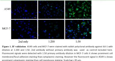
Highly expressed in heart followed by skeletal muscle, pancreas and kidney. Weakly expressed in placenta, lung and liver.

Post-translational modifications:
Autophosphorylated on serine residues.

Similarity:
Belongs to the protein kinase superfamily. TKL Ser/Thr protein kinase family.
Contains 5 ANK repeats.
Contains 1 protein kinase domain.

SWISS:
Q13418

Gene ID:
3611

Database links:

Entrez Gene: 3611 Human

Entrez Gene: 16202 Mouse

Entrez Gene: 170922 Rat

Omim: 602366 Human

SwissProt: Q13418 Human

SwissProt: O55222 Mouse

SwissProt: Q99J82 Rat

Unigene: 5158 Human

Unigene: 706355 Human

Unigene: 274846 Mouse

Unigene: 95042 Rat


ILK 是一种新发现的Ser/Thr蛋白激酶。ILK能够通过与整合素β1亚单位的结合介导细胞与胞外基质的连接,以依赖于PI3K的方式激活,并通过磷酸化下游底物PKB/AKT,GSK3等胞外信号的一项下游传递,对细胞的生长,分化,迁移等进行调控。由于ILK在胞内外信号传导中起着重要的作用。并且抑制ILK的活性能够导致细胞周期的停滞和细胞程序性死亡的启动,使其成为肿瘤治疗和肿瘤药物的理想靶位点。
该蛋白也表达与肾小球系膜细胞,正常足细胞ILK的高水平表达对足细胞功能起着重要的生理作用。DN 患者ILK表达明显增加 ,暴露于高血糖的肾小球膜细胞的ILK 水平明显升高, ILK 与整合素和其他连接蛋白都受到高血糖的调节,并且促进DN 患者肾小球基质的沉积。 Picture

Sample:
Lung (Mouse) Lysate at 40 ug
Heart (Mouse) Lysate at 40 ug
Primary: Anti-ILK-1 (SL0317R) at 1/300 dilution
Secondary: IRDye800CW Goat Anti-Rabbit IgG at 1/20000 dilution
Predicted band size: 50 kD
Observed band size: 50 kD
Sample:
Lane 1: Mouse Heart tissue lysates
Lane 2: Mouse Muscle tissue lysates
Lane 3: Rat Heart tissue lysates
Lane 4: Rat Muscle tissue lysates
Lane 5: Human MCF-7 cell lysates
Lane 6: Human Huvec cell lysates
Lane 7: Human U87MG cell lysates
Primary: Anti-ILK-1 (SL0317R) at 1/1000 dilution
Secondary: IRDye800CW Goat Anti-Rabbit IgG at 1/20000 dilution
Predicted band size: 50 kDa
Observed band size: 50 kDa
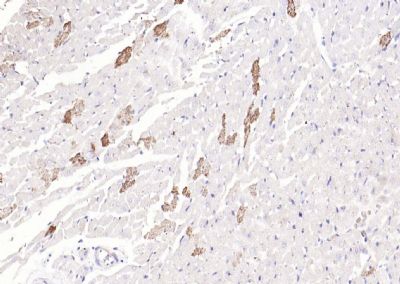
Paraformaldehyde-fixed, paraffin embedded (rat heart); Antigen retrieval by boiling in sodium citrate buffer (pH6.0) for 15min; Block endogenous peroxidase by 3% hydrogen peroxide for 20 minutes; Blocking buffer (normal goat serum) at 37°C for 30min; Antibody incubation with (ILK-1 ) Polyclonal Antibody, Unconjugated (SL0317R) at 1:200 overnight at 4°C, followed by operating according to SP Kit(Rabbit) (sp-0023) instructionsand DAB staining.
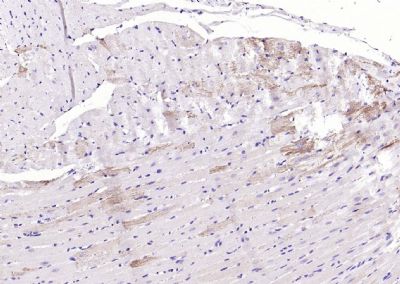
Paraformaldehyde-fixed, paraffin embedded (mouse heart); Antigen retrieval by boiling in sodium citrate buffer (pH6.0) for 15min; Block endogenous peroxidase by 3% hydrogen peroxide for 20 minutes; Blocking buffer (normal goat serum) at 37°C for 30min; Antibody incubation with (ILK-1 ) Polyclonal Antibody, Unconjugated (SL0317R) at 1:200 overnight at 4°C, followed by operating according to SP Kit(Rabbit) (sp-0023) instructionsand DAB staining.
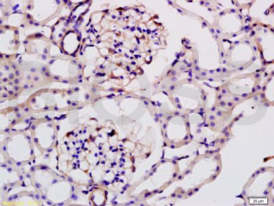
Tissue/cell: rat kidney tissue; 4% Paraformaldehyde-fixed and paraffin-embedded;
Antigen retrieval: citrate buffer ( 0.01M, pH 6.0 ), Boiling bathing for 15min; Block endogenous peroxidase by 3% Hydrogen peroxide for 30min; Blocking buffer (normal goat serum,SLC0005) at 37℃ for 20 min;
Incubation: Anti-ILK-1 Polyclonal Antibody, Unconjugated(SL0317R) 1:200, overnight at 4°C, followed by conjugation to the secondary antibody(SP-0023) and DAB(SLC0010) staining
Product Feedback Wall
Message :
Your Email :
Copyright © 2007-2018 Sunlong Medical All Rights Reserved.