Home > Product > Antibody > Rabbit Anti-Nogo-A antibody
Nogo A; reticulon-4 isoform D; ASY; Foocen; Human NogoA; KIAA0886; My043 protein; Nbla00271; Nbla10545; Neurite growth inhibitor 220; Neurite Outgrowth Inhibitor; Neuroendocrine specific protein; Neuroendocrine specific protein C homolog; NI220/250; Nogo
Cat:
SL0134R
Species Reactivity:
Human,Rat,(predicted: Mouse,)
Immunogen:
KLH conjugated synthetic peptide derived from mouse RTN4 protein:301-400/1192
Format:
Liquid
Storage instructions:
Shipped at 4℃. Store at -20 °C for one year. Avoid repeated freeze/thaw cycles.
Concentration:
1mg/ml
Clonality:
Polyclonal
Isotype:
IgG
Applications:
WB=1:500-2000ELISA=1:5000-10000IHC-P=1:100-500IHC-F=1:100-500Flow-Cyt=3μg/TestIF=1:100-500(Paraffin sections need to do antigen repair)not yet tested in other applications.optimal dilutions/concentrations should be determined by the end user.
Host:
Rabbit
Product Overview:
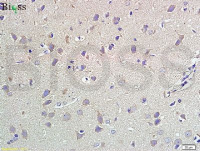
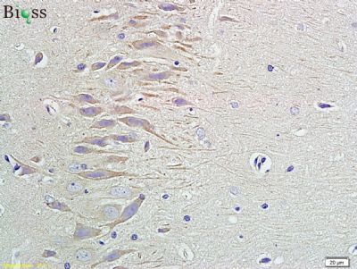
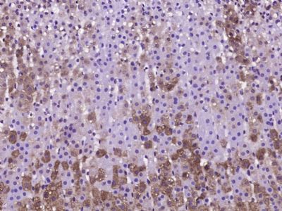
Tissue/cell: human meningioma tissue; 4% Paraformaldehyde-fixed and paraffin-embedded; Antigen retrieval: citrate buffer ( 0.01M, pH 6.0 ), Boiling bathing for 15min; Block endogenous peroxidase by 3% Hydrogen peroxide for 30min; Blocking buffer (normal goat serum,SLC0005) at 37℃ for 20 min; Incubation: Anti-Nogo-A Polyclonal Antibody, Unconjugated(SL0134R) 1:500, overnight at 4°C, followed by conjugation to the secondary antibody(SP-0023) and DAB(SLC0010) stainingParaformaldehyde-fixed, paraffin embedded (rat brain tissue); Antigen retrieval by boiling in sodium citrate buffer (pH6.0) for 15min; Block endogenous peroxidase by 3% hydrogen peroxide for 20 minutes; Blocking buffer (normal goat serum) at 37°C for 30min; Antibody incubation with (Nogo-A) Polyclonal Antibody, Unconjugated (SL0134R) at 1:400 overnight at 4°C, followed by operating according to SP Kit(Rabbit) (sp-0023) instructionsand DAB staining.Blank control: A431. Primary Antibody (green line): Rabbit Anti-Nogo-A antibody (SL0134R) Dilution: 1μg /10^6 cells; Isotype Control Antibody (orange line): Rabbit IgG . Secondary Antibody: Goat anti-rabbit IgG-AF647Dilution: 1μg /test. ProtocolThe cells were fixed with 4% PFA (10min at room temperature)and then permeabilized with 0.1% PBST for 20 min at room temperature. The cells were then incubated in 5%BSA to block non-specific protein-protein interactions for 30 min at room temperature.Cells stained with Primary Antibody for 30 min at room temperature. The secondary antibody used for 40 min at room temperature. Acquisition of 20,000 events was performed.
More
Unit:
Price: $
Product PDFs
Datasheet:


This gene belongs to the family of reticulon encoding genes. Reticulons are associated with the endoplasmic reticulum, and are involved in neuroendocrine secretion or in membrane trafficking in neuroendocrine cells. The product of this gene is a potent neurite outgrowth inhibitor which may also help block the regeneration of the central nervous system in higher vertebrates. Alternatively spliced transcript variants derived both from differential splicing and differential promoter usage and encoding different isoforms have been identified. [provided by RefSeq, Jul 2008].

Function:
Developmental neurite growth regulatory factor with a role as a negative regulator of axon-axon adhesion and growth, and as a facilitator of neurite branching. Regulates neurite fasciculation, branching and extension in the developing nervous system. Involved in down-regulation of growth, stabilization of wiring and restriction of plasticity in the adult CNS. Regulates the radial migration of cortical neurons via an RTN4R-LINGO1 containing receptor complex (By similarity). Isoform 2 reduces the anti-apoptotic activity of Bcl-xl and Bcl-2. This is likely consecutive to their change in subcellular location, from the mitochondria to the endoplasmic reticulum, after binding and sequestration. Isoform 2 and isoform 3 inhibit BACE1 activity and amyloid precursor protein processing.

Subunit:
Binds to RTN4R. Interacts with Bcl-xl and Bcl-2. Isoform 2 binds to NGBR and RTN3. Isoform 2 and isoform 3 interact with BACE1 and BACE2. Interacts with RTN4IP1. Interacts with ATL1.

Subcellular Location:
Endoplasmic reticulum membrane. Anchored to the membrane of the endoplasmic reticulum through 2 putative transmembrane domains.

Tissue Specificity:
Isoform 1 is specifically expressed in brain and testis and weakly in heart and skeletal muscle. Isoform 2 is widely expressed except for the liver. Isoform 3 is expressed in brain, skeletal muscle and adipocytes. Isoform 4 is testis-specific.

Similarity:
Contains 1 reticulon domain.

SWISS:
Q9NQC3

Gene ID:
57142

Database links:

Entrez Gene: 57142 Human

Entrez Gene: 68585 Mouse

Entrez Gene: 83765 Rat

Omim: 604475 Human

SwissProt: Q9NQC3 Human

SwissProt: Q99P72 Mouse

SwissProt: Q9JK11 Rat

Unigene: 704007 Human


神经生物学相关蛋白(Neurobiology)
Nogo-A轴索生长抑制因子在中枢神经的髓鞘上表达并在抑制损伤轴索修复中起着关键作用,从而有限制神经功能恢复的现象。主要定位细胞核。 Picture

Tissue/cell: human meningioma tissue; 4% Paraformaldehyde-fixed and paraffin-embedded;
Antigen retrieval: citrate buffer ( 0.01M, pH 6.0 ), Boiling bathing for 15min; Block endogenous peroxidase by 3% Hydrogen peroxide for 30min; Blocking buffer (normal goat serum,SLC0005) at 37℃ for 20 min;
Incubation: Anti-Nogo-A Polyclonal Antibody, Unconjugated(SL0134R) 1:500, overnight at 4°C, followed by conjugation to the secondary antibody(SP-0023) and DAB(SLC0010) staining
Paraformaldehyde-fixed, paraffin embedded (rat brain tissue); Antigen retrieval by boiling in sodium citrate buffer (pH6.0) for 15min; Block endogenous peroxidase by 3% hydrogen peroxide for 20 minutes; Blocking buffer (normal goat serum) at 37°C for 30min; Antibody incubation with (Nogo-A) Polyclonal Antibody, Unconjugated (SL0134R) at 1:400 overnight at 4°C, followed by operating according to SP Kit(Rabbit) (sp-0023) instructionsand DAB staining.
Blank control: A431.
Primary Antibody (green line): Rabbit Anti-Nogo-A antibody (SL0134R)
Dilution: 1μg /10^6 cells;
Isotype Control Antibody (orange line): Rabbit IgG .
Secondary Antibody: Goat anti-rabbit IgG-AF647
Dilution: 1μg /test.
Protocol
The cells were fixed with 4% PFA (10min at room temperature)and then permeabilized with 0.1% PBST for 20 min at room temperature. The cells were then incubated in 5%BSA to block non-specific protein-protein interactions for 30 min at room temperature.Cells stained with Primary Antibody for 30 min at room temperature. The secondary antibody used for 40 min at room temperature. Acquisition of 20,000 events was performed.
Product Feedback Wall
Message :
Your Email :
Copyright © 2007-2018 Sunlong Medical All Rights Reserved.