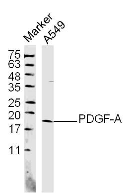
Home > Product > Antibody > Rabbit Anti-PDGF-A antibody
Platelet–dirived growth factor–1; PDGFA; PDGF AA; PDGF-AA; PDGF A chain; PDGF subunit A; PDGF-1; PDGF1; PDGFA; PDGFA_HUMAN; Platelet derived growth factor A chain precursor; Platelet derived growth factor alpha polypeptide; Platelet-derived growth factor
Cat:
SL0196R
Species Reactivity:
Human,Mouse,(predicted: Rat,Rabbit,)
Immunogen:
KLH conjugated synthetic peptide derived from the missle of human PDGF-A:125-170/211
Format:
Liquid
Storage instructions:
Shipped at 4℃. Store at -20 °C for one year. Avoid repeated freeze/thaw cycles.
Concentration:
1mg/ml
Clonality:
Polyclonal
Isotype:
IgG
Applications:
WB=1:500-2000ELISA=1:5000-10000IHC-P=1:100-500IHC-F=1:100-500Flow-Cyt=1μg/TestIF=1:100-500(Paraffin sections need to do antigen repair)not yet tested in other applications.optimal dilutions/concentrations should be determined by the end user.
Host:
Rabbit
Product Overview:
Sample: A549 (human)Cell Lysate at 40 ugPrimary: Anti- PDGF-A (SL0196R) at 1/300 dilutionSecondary: IRDye800CW Goat Anti-Rabbit IgG at 1/20000 dilutionPredicted band size: 14 kDObserved band size: 18 kDSample: placenta (Mouse) Lysate at 40 ugPrimary: Anti-PDGF-A (SL0196R) at 1/300 dilutionSecondary: IRDye800CW Goat Anti-Rabbit IgG at 1/20000 dilutionPredicted band size: 14 kDObserved band size: 17 kDBlank control (blue line): A431 cells (blue). Primary Antibody (green line): Rabbit Anti-PDGF-A antibody (SL0196R) Dilution: 1μg /10^6 cells; Isotype Control Antibody (orange line): Rabbit IgG . Secondary Antibody (white blue line): Goat anti-rabbit IgG-FITCDilution: 1μg /test. ProtocolThe cells were fixed with 70% methanol (Overnight at 4℃) and then permeabilized with 90% ice-cold methanol for 20 min at -20℃. Cells stained with Primary Antibody for 30 min at room temperature. The cells were then incubated in 1 X PBS/2%BSA/10% goat serum to block non-specific protein-protein interactions followed by the antibody for 15 min at room temperature. The secondary antibody used for 40 min at room temperature. Acquisition of 20,000 events was performed.
More
Unit:
Price: $
Product PDFs
Datasheet:


Platelet-derived growth factor is a potent mitogen for cells of mesenchymal origin. Binding of this growth factor to its affinity receptor elicits a variety of cellular responses. It is released by platelets upon wounding and plays an important role in stimulating adjacent cells to grow and thereby heal the wound. Antiparallel disulfide-linked dimer of nonidentical (A and B) chains. Homodimers of A and B chains are implicated in transformation processes. The long form contains a basic insert which acts as a cell retention signal. A-A and B-B, as well as A-B, dimers can bind to the PDGF receptor. It belongs to the PDGF/VEGF growth factor family.

Function:
Growth factor that plays an essential role in the regulation of embryonic development, cell proliferation, cell migration, survival and chemotaxis. Potent mitogen for cells of mesenchymal origin. Required for normal lung alveolar septum formation during embryogenesis, normal development of the gastrointestinal tract, normal development of Leydig cells and spermatogenesis. Required for normal oligodendrocyte development and normal myelination in the spinal cord and cerebellum. Plays an important role in wound healing. Signaling is modulated by the formation of heterodimers with PDGFB.

Subunit:
Homodimer; antiparallel disulfide-linked dimer. Heterodimer with PDGFB; antiparallel disulfide-linked dimer. The PDGFA homodimer interacts with PDGFRA homodimers, and with heterodimers formed by PDGFRA and PDGFRB. The heterodimer composed of PDGFA and PDGFB interacts with PDGFRA homodimers, and with heterodimers formed by PDGFRA and PDGFRB. Interacts with CSPG4.

Subcellular Location:
Secreted. Note=Released by platelets upon wounding.

Tissue Specificity:
Platelet-derived growth factor is a potent mitogen for cells of mesenchymal origin. Binding of this growth factor to its receptor elicits a variety of cellular responses. It is released by platelets upon wounding and plays an important role in stimulating adjacent cells to grow and thereby heals the wound.

Similarity:
Belongs to the PDGF/VEGF growth factor family.

SWISS:
P04085

Gene ID:
5154

Database links:

Entrez Gene: 5154 Human

Omim: 173430 Human

SwissProt: P04085 Human

Unigene: 535898 Human


PDGF-A(platelet-dirived growth factor-A)是一种碱性蛋白,pdgf促进细胞分裂、增值同时,可诱导c-fos、c-myc等蛋白表达增强。
主要作用:参与组织和血管修复、促进平滑肌细胞、成纤维细胞迅速增值。分子量为28-35kDa,分为两个亚基(A、B链)通过二硫键结合而成的两条多肽链的异二聚体。最新研究发现了PDGF家族两种新的基因PDGF-C、PDGF-D。 Picture

Sample: A549 (human)Cell Lysate at 40 ug
Primary: Anti- PDGF-A (SL0196R) at 1/300 dilution
Secondary: IRDye800CW Goat Anti-Rabbit IgG at 1/20000 dilution
Predicted band size: 14 kD
Observed band size: 18 kD
Sample:
placenta (Mouse) Lysate at 40 ug
Primary: Anti-PDGF-A (SL0196R) at 1/300 dilution
Secondary: IRDye800CW Goat Anti-Rabbit IgG at 1/20000 dilution
Predicted band size: 14 kD
Observed band size: 17 kD
Blank control (blue line): A431 cells (blue).
Primary Antibody (green line): Rabbit Anti-PDGF-A antibody (SL0196R)
Dilution: 1μg /10^6 cells;
Isotype Control Antibody (orange line): Rabbit IgG .
Secondary Antibody (white blue line): Goat anti-rabbit IgG-FITC
Dilution: 1μg /test.
Protocol
The cells were fixed with 70% methanol (Overnight at 4℃) and then permeabilized with 90% ice-cold methanol for 20 min at -20℃. Cells stained with Primary Antibody for 30 min at room temperature. The cells were then incubated in 1 X PBS/2%BSA/10% goat serum to block non-specific protein-protein interactions followed by the antibody for 15 min at room temperature. The secondary antibody used for 40 min at room temperature. Acquisition of 20,000 events was performed.
Product Feedback Wall
Message :
Your Email :
Copyright © 2007-2018 Sunlong Medical All Rights Reserved.