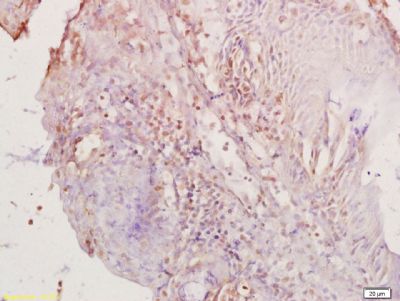
Tissue/cell: rat colon tissue; 4% Paraformaldehyde-fixed and paraffin-embedded;
Antigen retrieval: citrate buffer ( 0.01M, pH 6.0 ), Boiling bathing for 15min; Block endogenous peroxidase by 3% Hydrogen peroxide for 30min; Blocking buffer (normal goat serum,SLC0005) at 37℃ for 20 min;
Incubation: Anti-PAX8 Polyclonal Antibody, Unconjugated(SL1201R) 1:200, overnight at 4°C, followed by conjugation to the secondary antibody(SP-0023) and DAB(SLC0010) staining
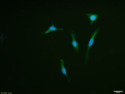
U87MG cell; 4% Paraformaldehyde-fixed; Triton X-100 at room temperature for 20 min; Blocking buffer (normal goat serum, SLC0005) at 37°C for 20 min; Antibody incubation with (PAX8) polyclonal Antibody, Unconjugated (SL1201R) 1:100, 90 minutes at 37°C; followed by a conjugated Goat Anti-Rabbit IgG antibody at 37°C for 90 minutes, DAPI (blue, C02-04002) was used to stain the cell nuclei.
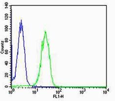
Cell:HT-29;
Concentration:1:100
Host/Isotype:Rabbit/IgG(blue line)
Flow cytometric analysis of Rabbit IgG isotype control on HT-29(blue) compared with primary antibody(Cat#: SL1021R)(green) followed by Alexa Fluor 488-conjugated goat anti-rabbit IgG(H+L) secondary antibody .
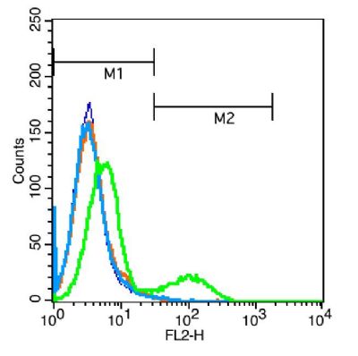
Primary Antibody:Rabbit Anti-TLR4 antibody(SL1021R, green line);
Isotype Control Antibody: Rabbit IgG (orange line);
Secondary Antibody: F(ab’)2 fragment goat anti-rabbit IgG-PE (white blue line)
Positive control: Jurkat cells(blue line)
Protocol
1.Jurkat cells were washed twice with phosphate-buffered saline (PBS).
2. An equivalent amount of pre-warmed 4% paraformaldehyde was added and the cells were incubated for 1 0 min at 37 °C.
|