Home > Product > Antibody > Rabbit Anti-PDGF-B antibody
PDGF beta; PDGF-B; Platelet-derived growth factor B-chain; PDGF AB + BB; Becaplermin; c sis; FLJ1285; Oncogene SIS; PDGF 1; PDGF 2; PDGF A; PDGF A chain; PDGF B chain; PDGF subunit A; PDGF subunit B; PDGF1; PDGF2; PDGFA; PDGFB; PDGFR1; Platelet derived gr
Cat:
SL0185R
Species Reactivity:
Human,Mouse,Rat,(predicted: Dog,Cow,Horse,Rabbit,Sheep,)
Immunogen:
KLH conjugated synthetic peptide derived from human PDGF-B:155-210/241
Format:
Liquid
Storage instructions:
Shipped at 4℃. Store at -20 °C for one year. Avoid repeated freeze/thaw cycles.
Concentration:
1mg/ml
Clonality:
Polyclonal
Isotype:
IgG
Applications:
WB=1:500-2000ELISA=1:5000-10000IHC-P=1:100-500IHC-F=1:100-500Flow-Cyt=1μg/TestIF=1:100-500(Paraffin sections need to do antigen repair)not yet tested in other applications.optimal dilutions/concentrations should be determined by the end user.
Host:
Rabbit
Product Overview:
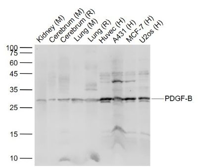
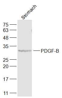
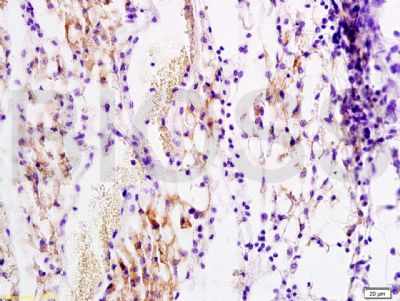
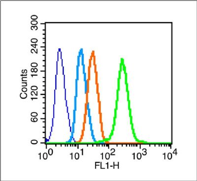
Sample: Lane 1: Kidney (Mouse) Lysate at 40 ugLane 2: Cerebrum (Mouse) Lysate at 40 ugLane 3: Cerebrum (Rat) Lysate at 40 ugLane 4: Lung (Mouse) Lysate at 40 ugLane 5: Lung (Rat) Lysate at 40 ugLane 6: Huvec (Human) Cell Lysate at 30 ugLane 7: A431 (Human) Cell Lysate at 30 ugLane 8: MCF-7 (Human) Cell Lysate at 30 ugLane 9: U2os (Human) Cell Lysate at 30 ugPrimary: Anti-PDGF-B (SL0185R) at 1/1000 dilutionSecondary: IRDye800CW Goat Anti-Rabbit IgG at 1/20000 dilutionPredicted band size: 25 kDObserved band size: 28 kDSample: Stomach (Rat) Lysate at 40 ugPrimary: Anti-PDGF-B (SL0185R) at 1/1000 dilutionSecondary: IRDye800CW Goat Anti-Rabbit IgG at 1/20000 dilutionPredicted band size: 27 kDObserved band size: 32 kDTissue/cell: rat transplant lymphoma; 4% Paraformaldehyde-fixed and paraffin-embedded; Antigen retrieval: citrate buffer ( 0.01M, pH 6.0 ), Boiling bathing for 15min; Block endogenous peroxidase by 3% Hydrogen peroxide for 30min; Blocking buffer (normal goat serum,SLC0005) at 37℃ for 20 min; Incubation: Anti-PDGF-B Polyclonal Antibody, Unconjugated(SL0071R) 1:200, overnight at 4°C, followed by conjugation to the secondary antibody(SP-0023) and DAB(SLC0010) stainingBlank control (blue line):Hela(blue). Primary Antibody (green line): Rabbit Anti-PDGF-B antibody (SL0185R) Dilution: 1μg /10^6 cells; Isotype Control Antibody (orange line): Rabbit IgG . Secondary Antibody (white blue line): F(ab’)2 fragment goat anti-rabbit IgG-FITCDilution: 1μg /test. ProtocolThe cells were fixed with 2% paraformaldehyde (10 min) , then permeabilized with 90% ice-cold methanol for 30 min on ice.Cells stained with Primary Antibody for 30 min at room temperature. The cells were then incubated in 1 X PBS/2%BSA/10% goat serum to block non-specific protein-protein interactions followed by the antibody for 15 min at room temperature. The secondary antibody used for 40 min at room temperature. Acquisition of 20,000 events was performed.
More
Unit:
Price: $
Product PDFs
Datasheet:


Platelet-derived growth factor is a potent mitogen for cells of mesenchymal origin. Binding of this growth factor to its affinity receptor elicits a variety of cellular responses. It is released by platelets upon wounding and plays an important role in stimulating adjacent cells to grow and thereby heal the wound. [SUBUNIT] Antiparallel disulfide-linked dimer of nonidentical (A and B) chains. Homodimers of A and B chains are implicated in transformation processes. A-A and B-B, as well as A-B, dimers can bind to the PDGF receptor. Belongs to the PDGF/VEGF growth factor family.

Function:
Growth factor that plays an essential role in the regulation of embryonic development, cell proliferation, cell migration, survival and chemotaxis. Potent mitogen for cells of mesenchymal origin. Required for normal proliferation and recruitment of pericytes and vascular smooth muscle cells in the central nervous system, skin, lung, heart and placenta. Required for normal blood vessel development, and for normal development of kidney glomeruli. Plays an important role in wound healing. Signaling is modulated by the formation of heterodimers with PDGFA.

Subunit:
Homodimer; antiparallel disulfide-linked dimer. Heterodimer with PDGFA; antiparallel disulfide-linked dimer. The PDGFB homodimer interacts with PDGFRA and PDGFRB homodimers, and with heterodimers formed by PDGFRA and PDGFRB. The heterodimer composed of PDGFA and PDGFB interacts with PDGFRB homodimers, and with heterodimers formed by PDGFRA and PDGFRB. Interacts with XLKD1.

Subcellular Location:
Secreted. Note=Released by platelets upon wounding.

Tissue Specificity:
Expressed at high levels in the heart, brain (sustantia nigra), placenta and fetal kidney. Expressed at moderate levels in the brain (hippocampus), skeletal muscle, kidney and lung.

DISEASE:
Note=A chromosomal aberration involving PDGFB is found in dermatofibrosarcoma protuberans. Translocation t(17;22)(q22;q13) with PDGFB.

Similarity:
Belongs to the PDGF/VEGF growth factor family.

SWISS:
P01127

Gene ID:
5155

Database links:

Entrez Gene: 5155 Human

Entrez Gene: 18591 Mouse

Entrez Gene: 24628 Rat

Omim: 190040 Human

SwissProt: P01127 Human

SwissProt: P3148 Mouse

SwissProt: Q05028 Rat

Unigene: 1976 Human

Unigene: 18889 Mouse

Unigene: 198230 Rat


生长因子和激素( Growth Factor and Hormones)
PDGF(platelet-dirived growth factor)是一种碱性蛋白,分为两个亚基(A、B链)通过二硫键结合而成的两条多肽链的异二聚体,分子量为28-35kDa。为促细胞分裂剂,可使小鼠成纤维细胞及其他多种细胞增殖。最新研究发现了PDGF家族两种新的基因PDGF-C、PDGF-D。
Picture

Sample:
Lane 1: Kidney (Mouse) Lysate at 40 ug
Lane 2: Cerebrum (Mouse) Lysate at 40 ug
Lane 3: Cerebrum (Rat) Lysate at 40 ug
Lane 4: Lung (Mouse) Lysate at 40 ug
Lane 5: Lung (Rat) Lysate at 40 ug
Lane 6: Huvec (Human) Cell Lysate at 30 ug
Lane 7: A431 (Human) Cell Lysate at 30 ug
Lane 8: MCF-7 (Human) Cell Lysate at 30 ug
Lane 9: U2os (Human) Cell Lysate at 30 ug
Primary: Anti-PDGF-B (SL0185R) at 1/1000 dilution
Secondary: IRDye800CW Goat Anti-Rabbit IgG at 1/20000 dilution
Predicted band size: 25 kD
Observed band size: 28 kD
Sample:
Stomach (Rat) Lysate at 40 ug
Primary: Anti-PDGF-B (SL0185R) at 1/1000 dilution
Secondary: IRDye800CW Goat Anti-Rabbit IgG at 1/20000 dilution
Predicted band size: 27 kD
Observed band size: 32 kD
Tissue/cell: rat transplant lymphoma; 4% Paraformaldehyde-fixed and paraffin-embedded;
Antigen retrieval: citrate buffer ( 0.01M, pH 6.0 ), Boiling bathing for 15min; Block endogenous peroxidase by 3% Hydrogen peroxide for 30min; Blocking buffer (normal goat serum,SLC0005) at 37℃ for 20 min;
Incubation: Anti-PDGF-B Polyclonal Antibody, Unconjugated(SL0071R) 1:200, overnight at 4°C, followed by conjugation to the secondary antibody(SP-0023) and DAB(SLC0010) staining
Blank control (blue line):Hela(blue).
Primary Antibody (green line): Rabbit Anti-PDGF-B antibody (SL0185R)
Dilution: 1μg /10^6 cells;
Isotype Control Antibody (orange line): Rabbit IgG .
Secondary Antibody (white blue line): F(ab’)2 fragment goat anti-rabbit IgG-FITC
Dilution: 1μg /test.
Protocol
The cells were fixed with 2% paraformaldehyde (10 min) , then permeabilized with 90% ice-cold methanol for 30 min on ice.Cells stained with Primary Antibody for 30 min at room temperature. The cells were then incubated in 1 X PBS/2%BSA/10% goat serum to block non-specific protein-protein interactions followed by the antibody for 15 min at room temperature. The secondary antibody used for 40 min at room temperature. Acquisition of 20,000 events was performed.
Product Feedback Wall
Message :
Your Email :
Copyright © 2007-2018 Sunlong Medical All Rights Reserved.