Home > Product > Antibody > Rabbit Anti-CD166 antibody
activated leukocyte cell adhesion molecule; ALCAM protein; ALCAM; CD 166; CD166 antigen; FLJ38514; MEMD; MGC71733; FLJ3851; AI853494; BEN; DM-GRASP; MGC27910; MuSC; SC1; CD166_HUMAN.
Cat:
SL1251R
Species Reactivity:
Human,(predicted: Mouse,Rat,Chicken,Dog,Pig,Cow,Horse,Rabbit,)
Immunogen:
KLH conjugated synthetic peptide derived from human CD166:451-583/583<Cytoplasmic>
Format:
Liquid
Storage instructions:
Shipped at 4℃. Store at -20 °C for one year. Avoid repeated freeze/thaw cycles.
Concentration:
1mg/ml
Clonality:
Polyclonal
Isotype:
IgG
Applications:
WB=1:500-2000ELISA=1:5000-10000IHC-F=1:100-500Flow-Cyt=1μg/TestIF=1:100-500(Paraffin sections need to do antigen repair)not yet tested in other applications.optimal dilutions/concentrations should be determined by the end user.
Host:
Rabbit
Product Overview:
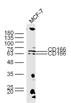
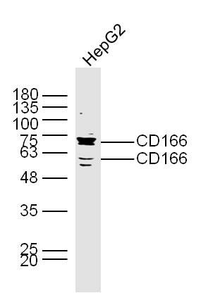
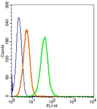
Sample: MCF-7 Cell (Human) Lysate at 30 ugPrimary: Anti-CD166 (SL1251R) at 1/300 dilutionSecondary: IRDye800CW Goat Anti-Rabbit IgG at 1/20000 dilutionPredicted band size: 62 kDObserved band size: 62/70 kDSample: HepG2 Cell (Human) Lysate at 30 ugPrimary: Anti-CD166 (SL1251R) at 1/300 dilutionSecondary: IRDye800CW Goat Anti-Rabbit IgG at 1/20000 dilutionPredicted band size: 62 kDObserved band size: 62/70 kDBlank control: Molt-4 Cells(blue). Primary Antibody: Rabbit Anti-CD166/FITC Conjugated antibody (SL1251R/AF488), Dilution: 1μg in 100 μL 1X PBS containing 0.5% BSA; Isotype Control Antibody: Rabbit IgG/AF488orange) ,used under the same conditions.ProtocolThe cells were fixed with 2% paraformaldehyde (10 min) . The cells were washed twice with 1 X PBS. The cells were incubated in 1 X PBS containing 0.5% BSA + 1 0% goat serum (15 min) to block non-specific protein-protein interactions followed by the incubated with antibody (SL1251R/AF488, 1μg /1x10^6 cells) for 30 min on ice. Acquisition of 20,000 events was performed.
More
Unit:
Price: $
Product PDFs
Datasheet:


CD166 is a member of the Ig superfamily and is expressed on activated T-cells, B cells and other cells including thymic epithelial cells, fibroblasts, keratinocytes and neurons. CD6 has been identified as a receptor for CD166.
The expression of CD166 is up-regulated in low-grade prostate tumors and down-regulated in high-grade tumors; may play role in progression of prostate cancer.

Function:
Cell adhesion molecule that binds to CD6. Involved in neurite extension by neurons via heterophilic and hemophilic interactions. May play a role in the binding of T- and B-cells to activated leukocytes, as well as in interactions between cells of the nervous system.

Subcellular Location:
Membrane; Single-pass type I membrane protein.

Tissue Specificity:
Spleen, placenta, liver, and weakly in liver. Expressed by activated T-cells, B-cells, monocytes and thymic epithelial cells. Expressed by neurons in the brain. Restricted expression in tumor cell lines. Preferentially expressed in highly metastasizing melanoma cell lines.

Similarity:
Contains 3 Ig-like C2-type (immunoglobulin-like) domains.
Contains 2 Ig-like SLVtype (immunoglobulin-like) domains.

SWISS:
Q13740

Gene ID:
214

Database links:

Entrez Gene: 214 Human

Entrez Gene: 11658 Mouse

Entrez Gene: 79559 Rat

Omim: 601662 Human

SwissProt: Q13740 Human

SwissProt: Q61490 Mouse

SwissProt: O35112 Rat

Unigene: 591293 Human

Unigene: 288282 Mouse

Unigene: 5789 Rat


ALCAM/CD166是免疫球蛋白超家族的成员,对多种肿瘤生长和转移相关的肿瘤细胞特性具有调节作用,ALCAM/CD166在乳腺癌、前列腺癌、食道癌、结肠癌及恶性黑色素瘤等肿瘤都有一定的表达,尤以结肠、直肠癌表达显著。 Picture

Sample:
MCF-7 Cell (Human) Lysate at 30 ug
Primary: Anti-CD166 (SL1251R) at 1/300 dilution
Secondary: IRDye800CW Goat Anti-Rabbit IgG at 1/20000 dilution
Predicted band size: 62 kD
Observed band size: 62/70 kD
Sample:
HepG2 Cell (Human) Lysate at 30 ug
Primary: Anti-CD166 (SL1251R) at 1/300 dilution
Secondary: IRDye800CW Goat Anti-Rabbit IgG at 1/20000 dilution
Predicted band size: 62 kD
Observed band size: 62/70 kD
Blank control: Molt-4 Cells(blue).
Primary Antibody: Rabbit Anti-CD166/FITC Conjugated antibody (SL1251R/AF488), Dilution: 1μg in 100 μL 1X PBS containing 0.5% BSA;
Isotype Control Antibody: Rabbit IgG/AF488orange) ,used under the same conditions.
Protocol
The cells were fixed with 2% paraformaldehyde (10 min) . The cells were washed twice with 1 X PBS. The cells were incubated in 1 X PBS containing 0.5% BSA + 1 0% goat serum (15 min) to block non-specific protein-protein interactions followed by the incubated with antibody (SL1251R/AF488, 1μg /1x10^6 cells) for 30 min on ice. Acquisition of 20,000 events was performed.
Product Feedback Wall
Message :
Your Email :
Copyright © 2007-2018 Sunlong Medical All Rights Reserved.