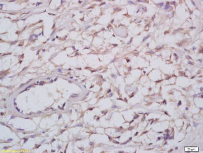
Tissue/cell: human cervical carcinoma; 4% Paraformaldehyde-fixed and paraffin-embedded;
Antigen retrieval: citrate buffer ( 0.01M, pH 6.0 ), Boiling bathing for 15min; Block endogenous peroxidase by 3% Hydrogen peroxide for 30min; Blocking buffer (normal goat serum,SLC0005) at 37℃ for 20 min;
Incubation: Anti-Nogo-B Polyclonal Antibody, Unconjugated(SL1315R) 1:200, overnight at 4°C, followed by conjugation to the secondary antibody(SP-0023) and DAB(SLC0010) staining
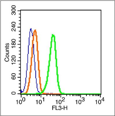
Blank control (blue line): A431(blue).
Primary Antibody (green line): Rabbit Anti-Nogo A+B/PE-CY5 Conjugated antibody (SL1315R-PE-CY5)
Dilution: 1μg /10^6 cells;
Isotype Control Antibody (orange line): Rabbit IgG-PE-CY5 .
Protocol
The cells were fixed with 70% ice-cold methanol overnight at 4℃ . The cells were then incubated in 1 X PBS/2%BSA/10% goat serum to block non-specific protein-protein interactions followed by the antibody for 15 min at room temperature. Cells stained with Primary Antibody for 30 min at room temperature.Acquisition of 20,000 events was performed.