Home > Product > Antibody > Rabbit Anti-Thioredoxin antibody
ADF; Surface associated sulphydryl protein; Thioredoxin-1; Thioredoxin 1; Thioredoxin1; TXN protein; ADF; ATL derived factor; DKFZp686B1993; MGC61975; SASP; Surface associated sulphydryl protein; Surface-associated sulphydryl protein; Thioredoxin; TRDX; T
Cat:
SL0458R
Species Reactivity:
Recombinant protein
Immunogen:
KLH conjugated synthetic peptide derived from human Thioredoxin:51-105/105
Format:
Liquid
Storage instructions:
Shipped at 4℃. Store at -20 °C for one year. Avoid repeated freeze/thaw cycles.
Concentration:
1mg/ml
Clonality:
Polyclonal
Isotype:
IgG
Applications:
WB=1:1000-5000ELISA=1:5000-10000IHC-P=1:100-500IHC-F=1:100-500Flow-Cyt=1µg/TestICC=1:100IF=1:100-500(Paraffin sections need to do antigen repair)not yet tested in other applications.optimal dilutions/concentrations should be determined by the end user.
Host:
Rabbit
Product Overview:
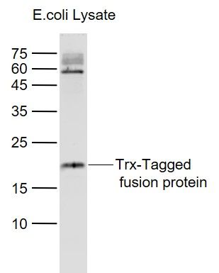
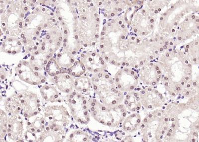
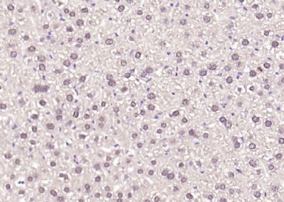
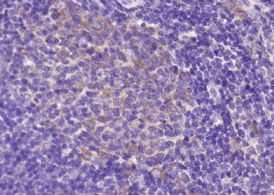
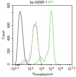
Sample: Lane 1: Trx-Tagged fusion protein (full length) Overexpression E.coli Lysate (Cat#: SL33018P) at 4 ug Primary: Anti-Thioredoxin/Trx (SL0458R) at 1/1000 dilution Secondary: IRDye800CW Goat Anti-Rabbit IgG at 1/20000 dilution Predicted band size: 18 kD Observed band size: 18 kDParaformaldehyde-fixed, paraffin embedded (mouse kidney); Antigen retrieval by boiling in sodium citrate buffer (pH6.0) for 15min; Block endogenous peroxidase by 3% hydrogen peroxide for 20 minutes; Blocking buffer (normal goat serum) at 37°C for 30min; Antibody incubation with (Thioredoxin) Polyclonal Antibody, Unconjugated (SL0458R) at 1:200 overnight at 4°C, followed by operating according to SP Kit(Rabbit) (sp-0023) instructionsand DAB staining.Paraformaldehyde-fixed, paraffin embedded (mouse liver); Antigen retrieval by boiling in sodium citrate buffer (pH6.0) for 15min; Block endogenous peroxidase by 3% hydrogen peroxide for 20 minutes; Blocking buffer (normal goat serum) at 37°C for 30min; Antibody incubation with (Thioredoxin) Polyclonal Antibody, Unconjugated (SL0458R) at 1:200 overnight at 4°C, followed by operating according to SP Kit(Rabbit) (sp-0023) instructionsand DAB staining.Paraformaldehyde-fixed, paraffin embedded (rat spleen); Antigen retrieval by boiling in sodium citrate buffer (pH6.0) for 15min; Block endogenous peroxidase by 3% hydrogen peroxide for 20 minutes; Blocking buffer (normal goat serum) at 37°C for 30min; Antibody incubation with (Thioredoxin) Polyclonal Antibody, Unconjugated (SL0458R) at 1:200 overnight at 4°C, followed by operating according to SP Kit(Rabbit) (sp-0023) instructionsand DAB staining.Paraformaldehyde-fixed, paraffin embedded (human kidney); Antigen retrieval by boiling in sodium citrate buffer (pH6.0) for 15min; Block endogenous peroxidase by 3% hydrogen peroxide for 20 minutes; Blocking buffer (normal goat serum) at 37°C for 30min; Antibody incubation with (Thioredoxin) Polyclonal Antibody, Unconjugated (SL0458R) at 1:400 overnight at 4°C, followed by a conjugated secondary (sp-0023) for 20 minutes and DAB staining. Paraformaldehyde-fixed, paraffin embedded (human cervical carcinoma); Antigen retrieval by boiling in sodium citrate buffer (pH6.0) for 15min; Block endogenous peroxidase by 3% hydrogen peroxide for 20 minutes; Blocking buffer (normal goat serum) at 37°C for 30min; Antibody incubation with (Thioredoxin) Polyclonal Antibody, Unconjugated (SL0458R) at 1:400 overnight at 4°C, followed by a conjugated secondary (sp-0023) for 20 minutes and DAB staining. Paraformaldehyde-fixed, paraffin embedded (rat brain); Antigen retrieval by boiling in sodium citrate buffer (pH6.0) for 15min; Block endogenous peroxidase by 3% hydrogen peroxide for 20 minutes; Blocking buffer (normal goat serum) at 37°C for 30min; Antibody incubation with (Thioredoxin) Polyclonal Antibody, Unconjugated (SL0458R) at 1:400 overnight at 4°C, followed by a conjugated secondary (sp-0023) for 20 minutes and DAB staining. Hela cell; 4% Paraformaldehyde-fixed; Triton X-100 at room temperature for 20 min; Blocking buffer (normal goat serum, SLC0005) at 37°C for 20 min; Antibody incubation with (Thioredoxin) polyclonal Antibody, Unconjugated (SL0458R) 1:100, 90 minutes at 37°C; followed by a conjugated Goat Anti-Rabbit IgG antibody at 37°C for 90 minutes, DAPI (blue, C02-04002) was used to stain the cell nuclei.Blank control: SHSY5Y. Primary Antibody (green line): Rabbit Anti-Thioredoxin antibody (SL0458R) Dilution: 1ug/Test; Secondary Antibody : Goat anti-rabbit IgG-FITCDilution: 0.5ug/Test. ProtocolThe cells were fixed with 4% PFA (10min at room temperature)and then permeabilized with 90% ice-cold methanol for 20 min at -20℃.The cells were then incubated in 5%BSA to block non-specific protein-protein interactions for 30 min at room temperature .Cells stained with Primary Antibody for 30 min at room temperature. The secondary antibody used for 40 min at room temperature. Acquisition of 20,000 events was performed.
More
Unit:
Price: $
Product PDFs
Datasheet:


The protein encoded by this gene acts as a homodimer and is involved in many redox reactions. The encoded protein is active in the reversible S-nitrosylation of cysteines in certain proteins, which is part of the response to intracellular nitric oxide. This protein is found in the cytoplasm. Two transcript variants encoding different isoforms have been found for this gene. [provided by RefSeq, Oct 2011]

Function:
Participates in various redox reactions through the reversible oxidation of its active center dithiol to a disulfide and catalyzes dithiol-disulfide exchange reactions. Plays a role in the reversible S-nitrosylation of cysteine residues in target proteins, and thereby contributes to the response to intracellular nitric oxide. Nitrosylates the active site Cys of CASP3 in response to nitric oxide (NO), and thereby inhibits caspase-3 activity. Induces the FOS/JUN AP-1 DNA-binding activity in ionizing radiation (IR) cells through its oxidation/reduction status and stimulates AP-1 transcriptional activity.

Subunit:
Homodimer; disulfide-linked. Interacts with TXNIP through the redox-active site. Interacts with MAP3K5 and CASP3. In case of infection, interacts with S.typhimurium protein slrP. Interacts with APEX1; the interaction stimulates the FOS/JUN AP-1 DNA-binding activity in a redox-dependent manner.

Subcellular Location:
Nucleus. Cytoplasm. Secreted. Note=Secreted by a leaderless secretory pathway. Predominantly in the cytoplasm in non irradiated cells. Radiation induces translocation of TRX from the cytoplasm to the nucleus.

Similarity:
Belongs to the thioredoxin family. Contains 1 thioredoxin domain.

SWISS:
P10599

Gene ID:
7295

Database links:

Entrez Gene: 7295 Human

Entrez Gene: 22166 Mouse

Entrez Gene: 116484 Rat

Omim: 187700 Human

SwissProt: P10599 Human

SwissProt: P10639 Mouse

SwissProt: P11232 Rat

Unigene: 435136 Human

Unigene: 260618 Mouse

Unigene: 29777 Rat


硫氧还蛋白(Thioredoxin,Trx)是一类广泛存在于生物体内的多功能酸性蛋白,分子量约12kDa,是一种小分子含硒蛋白质,参与细胞的一系列生化反应,包括酶活性的调节、转录因子的调控等,是重要的酶活力调节蛋白。
硫氧还蛋白的功能绝大多数是依赖于硫氧还蛋白还原靶蛋白中的二硫键。所有硫氧还蛋白均有一个保守的活性中心,Trp-Cys-Gly(Ala)-Pro-Cys,活性中心有两个具有氧还活性的半胱氨酸残基。 Picture

Sample:
Lane 1: Trx-Tagged fusion protein (full length) Overexpression E.coli Lysate (Cat#: SL33018P) at 4 ug
Primary: Anti-Thioredoxin/Trx (SL0458R) at 1/1000 dilution
Secondary: IRDye800CW Goat Anti-Rabbit IgG at 1/20000 dilution
Predicted band size: 18 kD
Observed band size: 18 kD
Paraformaldehyde-fixed, paraffin embedded (mouse kidney); Antigen retrieval by boiling in sodium citrate buffer (pH6.0) for 15min; Block endogenous peroxidase by 3% hydrogen peroxide for 20 minutes; Blocking buffer (normal goat serum) at 37°C for 30min; Antibody incubation with (Thioredoxin) Polyclonal Antibody, Unconjugated (SL0458R) at 1:200 overnight at 4°C, followed by operating according to SP Kit(Rabbit) (sp-0023) instructionsand DAB staining.
Paraformaldehyde-fixed, paraffin embedded (mouse liver); Antigen retrieval by boiling in sodium citrate buffer (pH6.0) for 15min; Block endogenous peroxidase by 3% hydrogen peroxide for 20 minutes; Blocking buffer (normal goat serum) at 37°C for 30min; Antibody incubation with (Thioredoxin) Polyclonal Antibody, Unconjugated (SL0458R) at 1:200 overnight at 4°C, followed by operating according to SP Kit(Rabbit) (sp-0023) instructionsand DAB staining.
Paraformaldehyde-fixed, paraffin embedded (rat spleen); Antigen retrieval by boiling in sodium citrate buffer (pH6.0) for 15min; Block endogenous peroxidase by 3% hydrogen peroxide for 20 minutes; Blocking buffer (normal goat serum) at 37°C for 30min; Antibody incubation with (Thioredoxin) Polyclonal Antibody, Unconjugated (SL0458R) at 1:200 overnight at 4°C, followed by operating according to SP Kit(Rabbit) (sp-0023) instructionsand DAB staining.
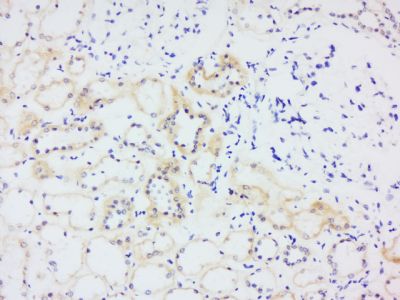
Paraformaldehyde-fixed, paraffin embedded (human kidney); Antigen retrieval by boiling in sodium citrate buffer (pH6.0) for 15min; Block endogenous peroxidase by 3% hydrogen peroxide for 20 minutes; Blocking buffer (normal goat serum) at 37°C for 30min; Antibody incubation with (Thioredoxin) Polyclonal Antibody, Unconjugated (SL0458R) at 1:400 overnight at 4°C, followed by a conjugated secondary (sp-0023) for 20 minutes and DAB staining.
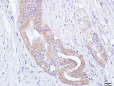
Paraformaldehyde-fixed, paraffin embedded (human cervical carcinoma); Antigen retrieval by boiling in sodium citrate buffer (pH6.0) for 15min; Block endogenous peroxidase by 3% hydrogen peroxide for 20 minutes; Blocking buffer (normal goat serum) at 37°C for 30min; Antibody incubation with (Thioredoxin) Polyclonal Antibody, Unconjugated (SL0458R) at 1:400 overnight at 4°C, followed by a conjugated secondary (sp-0023) for 20 minutes and DAB staining.
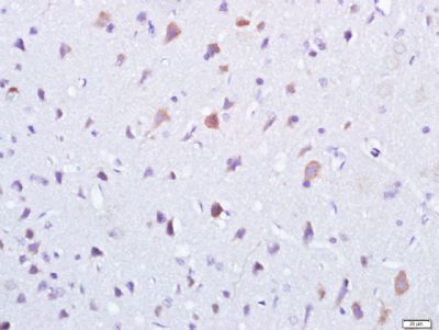
Paraformaldehyde-fixed, paraffin embedded (rat brain); Antigen retrieval by boiling in sodium citrate buffer (pH6.0) for 15min; Block endogenous peroxidase by 3% hydrogen peroxide for 20 minutes; Blocking buffer (normal goat serum) at 37°C for 30min; Antibody incubation with (Thioredoxin) Polyclonal Antibody, Unconjugated (SL0458R) at 1:400 overnight at 4°C, followed by a conjugated secondary (sp-0023) for 20 minutes and DAB staining.
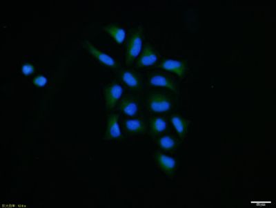
Hela cell; 4% Paraformaldehyde-fixed; Triton X-100 at room temperature for 20 min; Blocking buffer (normal goat serum, SLC0005) at 37°C for 20 min; Antibody incubation with (Thioredoxin) polyclonal Antibody, Unconjugated (SL0458R) 1:100, 90 minutes at 37°C; followed by a conjugated Goat Anti-Rabbit IgG antibody at 37°C for 90 minutes, DAPI (blue, C02-04002) was used to stain the cell nuclei.
Blank control: SHSY5Y.
Primary Antibody (green line): Rabbit Anti-Thioredoxin antibody (SL0458R)
Dilution: 1ug/Test;
Secondary Antibody : Goat anti-rabbit IgG-FITC
Dilution: 0.5ug/Test.
Protocol
The cells were fixed with 4% PFA (10min at room temperature)and then permeabilized with 90% ice-cold methanol for 20 min at -20℃.The cells were then incubated in 5%BSA to block non-specific protein-protein interactions for 30 min at room temperature .Cells stained with Primary Antibody for 30 min at room temperature. The secondary antibody used for 40 min at room temperature. Acquisition of 20,000 events was performed.
Product Feedback Wall
Message :
Your Email :
Copyright © 2007-2018 Sunlong Medical All Rights Reserved.