Home > Product > Antibody > Rabbit Anti-IGFBP3 antibody
IBP3_HUMAN; Insulin-like growth factor-binding protein 3; IBP3; IBP-3; IGF-binding protein 3; IGFBP-3; IBP3; BP-53;
Cat:
SL1434R
Species Reactivity:
Human,Rat,(predicted: Mouse,)
Immunogen:
KLH conjugated synthetic peptide derived from human IGFBP3:186-292/292
Format:
Liquid
Storage instructions:
Shipped at 4℃. Store at -20 °C for one year. Avoid repeated freeze/thaw cycles.
Concentration:
1mg/ml
Clonality:
Polyclonal
Isotype:
IgG
Applications:
WB=1:500-2000ELISA=1:5000-10000IHC-P=1:100-500IHC-F=1:100-500IF=1:100-500(Paraffin sections need to do antigen repair)not yet tested in other applications.optimal dilutions/concentrations should be determined by the end user.
Host:
Rabbit
Product Overview:
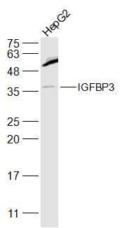
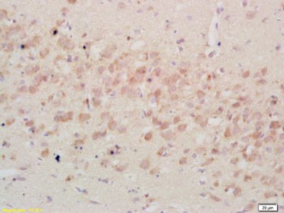
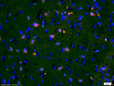
Sample: HepG2(Human) Cell Lysate at 30 ugPrimary: Anti- IGFBP3 (SL1434R) at 1/500 dilutionSecondary: IRDye800CW Goat Anti-Rabbit IgG at 1/20000 dilutionPredicted band size: 30-35 kDObserved band size: 40 kDTissue/cell: rat brain tissue; 4% Paraformaldehyde-fixed and paraffin-embedded; Antigen retrieval: citrate buffer ( 0.01M, pH 6.0 ), Boiling bathing for 15min; Block endogenous peroxidase by 3% Hydrogen peroxide for 30min; Blocking buffer (normal goat serum,SLC0005) at 37℃ for 20 min; Incubation: Anti-IGFBP3 Polyclonal Antibody, Unconjugated(SL1434R) 1:200, overnight at 4°C, followed by conjugation to the secondary antibody(SP-0023) and DAB(SLC0010) stainingTissue/cell: rat brain tissue;4% Paraformaldehyde-fixed and paraffin-embedded; Antigen retrieval: citrate buffer ( 0.01M, pH 6.0 ), Boiling bathing for 15min; Blocking buffer (normal goat serum,SLC0005) at 37℃ for 20 min; Incubation: Anti-IGFBP3 Polyclonal Antibody, Unconjugated(SL1434R) 1:200, overnight at 4°C; The secondary antibody was Goat Anti-Rabbit IgG, Cy3 conjugated(SL0295G-Cy3)used at 1:200 dilution for 40 minutes at 37°C. DAPI(5ug/ml,blue,SLC0033) was used to stain the cell nuclei
More
Unit:
Price: $
Product PDFs
Datasheet:


This gene is a member of the insulin-like growth factor binding protein (IGFBP) family and encodes a protein with an IGFBP domain and a thyroglobulin type-I domain. The protein forms a ternary complex with insulin-like growth factor acid-labile subunit (IGFALS) and either insulin-like growth factor (IGF) I or II. In this form, it circulates in the plasma, prolonging the half-life of IGFs and altering their interaction with cell surface receptors. Alternate transcriptional splice variants, encoding different isoforms, have been characterized. [provided by RefSeq, Jul 2008]

Subcellular Location:
Secreted.

Tissue Specificity:
Expressed by most tissues. Present in plasma.

Post-translational modifications:
Phosphorylation sites are present in the extracelllular medium.

Similarity:
Contains 1 IGFBP N-terminal domain.
Contains 1 thyroglobulin type-1 domain.

SWISS:
P17936

Gene ID:
3486

Database links:

Entrez Gene: 3486 Human

Entrez Gene: 16009 Mouse

Entrez Gene: 24484 Rat

SwissProt: P17936 Human

SwissProt: P47878 Mouse

SwissProt: P15473 Rat


Picture

Sample:
HepG2(Human) Cell Lysate at 30 ug
Primary: Anti- IGFBP3 (SL1434R) at 1/500 dilution
Secondary: IRDye800CW Goat Anti-Rabbit IgG at 1/20000 dilution
Predicted band size: 30-35 kD
Observed band size: 40 kD
Tissue/cell: rat brain tissue; 4% Paraformaldehyde-fixed and paraffin-embedded;
Antigen retrieval: citrate buffer ( 0.01M, pH 6.0 ), Boiling bathing for 15min; Block endogenous peroxidase by 3% Hydrogen peroxide for 30min; Blocking buffer (normal goat serum,SLC0005) at 37℃ for 20 min;
Incubation: Anti-IGFBP3 Polyclonal Antibody, Unconjugated(SL1434R) 1:200, overnight at 4°C, followed by conjugation to the secondary antibody(SP-0023) and DAB(SLC0010) staining
Tissue/cell: rat brain tissue;4% Paraformaldehyde-fixed and paraffin-embedded;
Antigen retrieval: citrate buffer ( 0.01M, pH 6.0 ), Boiling bathing for 15min; Blocking buffer (normal goat serum,SLC0005) at 37℃ for 20 min;
Incubation: Anti-IGFBP3 Polyclonal Antibody, Unconjugated(SL1434R) 1:200, overnight at 4°C; The secondary antibody was Goat Anti-Rabbit IgG, Cy3 conjugated(SL0295G-Cy3)used at 1:200 dilution for 40 minutes at 37°C. DAPI(5ug/ml,blue,SLC0033) was used to stain the cell nuclei
Product Feedback Wall
Message :
Your Email :
Copyright © 2007-2018 Sunlong Medical All Rights Reserved.